

Surgical Resection of an Optic Nerve Sheath Meningioma: Relevance of Endoscopic Endonasal Approaches to the Optic Canal
- PMID: 28413768
- PMCID: PMC5391263
- DOI: 10.1055/s-0037-1600897
Surgical Resection of an Optic Nerve Sheath Meningioma: Relevance of Endoscopic Endonasal Approaches to the Optic Canal
Abstract
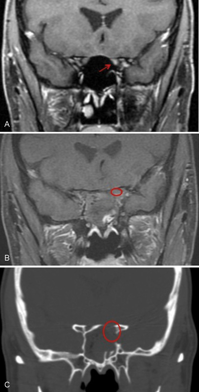
Optic nerve sheath meningiomas (ONSMs) account for less than 2% of meningiomas and 1.7% of orbital tumors. Although rare, the management of these tumors is important as unilateral blindness often results in untreated cases. Radiotherapy has emerged as the preferred treatment. However, therapies for ONSMs are controversial due to the variable natural history of the disease and limitations of surgical and radiotherapy options. A 60-year-old woman presented with monocular left diminished color perception and blurred vision. Magnetic resonance imaging demonstrated a homogenously enhancing 5-mm left optic nerve mass with evidence of nerve compression. Conservative management was advised. However, 1 month after diagnosis her visual acuity deteriorated further. Because of the small focal location of the tumor within the optic canal, surgery was considered. Given the tumor's location inferomedial to the optic nerve, an endoscopic endonasal approach to the optic canal was performed. This patient recovered fully with resolution of visual symptoms immediately following surgery. Postoperative imaging 24 hours after surgery demonstrated gross total resection of the tumor; 1 year postoperatively the patient has a normal ophthalmologic examination. This report highlights the value of endoscopic endonasal approaches in the management of select optic canal pathology, otherwise inaccessible via transcranial approaches.
Keywords: endoscopic endonasal; meningioma; minimally invasive; optic canal; optic nerve sheath; skull base.
Conflict of interest statement
Figures


References
-
- Castel A, Boschi A, Renard L, De Potter P. Optic nerve sheath meningiomas: clinical features, functional prognosis and controversial treatment. Bull Soc Belge Ophtalmol. 2000;275:73–78. - PubMed
-
- Dutton J J. Optic nerve sheath meningiomas. Surv Ophthalmol. 1992;37(03):167–183. - PubMed
-
- Shapey J, Sabin H I, Danesh-Meyer H V, Kaye A H. Diagnosis and management of optic nerve sheath meningiomas. J Clin Neurosci. 2013;20(08):1045–1056. - PubMed
-
- Abouaf L, Girard N, Lefort T et al.Standard-fractionated radiotherapy for optic nerve sheath meningioma: visual outcome is predicted by mean eye dose. Int J Radiat Oncol Biol Phys. 2012;82(03):1268–1277. - PubMed
-
- Becker G, Jeremic B, Pitz S et al.Stereotactic fractionated radiotherapy in patients with optic nerve sheath meningioma. Int J Radiat Oncol Biol Phys. 2002;54(05):1422–1429. - PubMed
Publication types
LinkOut - more resources
Full Text Sources
Other Literature Sources