. 2017 Mar 31:14:760-774.
doi: 10.1016/j.nicl.2017.03.016. eCollection 2017.

Family history and APOE4 risk for Alzheimer's disease impact the neural correlates of episodic memory by early midlife

Affiliations

Family history and APOE4 risk for Alzheimer's disease impact the neural correlates of episodic memory by early midlife

M N Rajah et al. Neuroimage Clin. .

Abstract

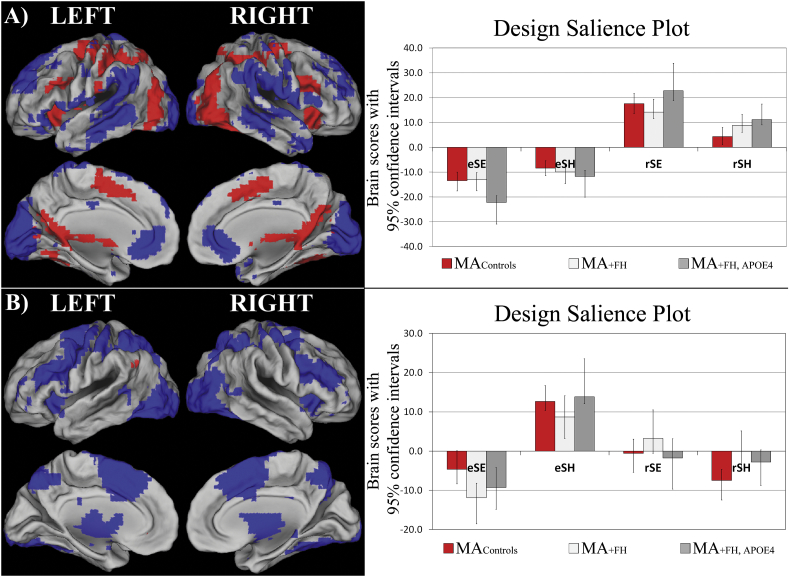
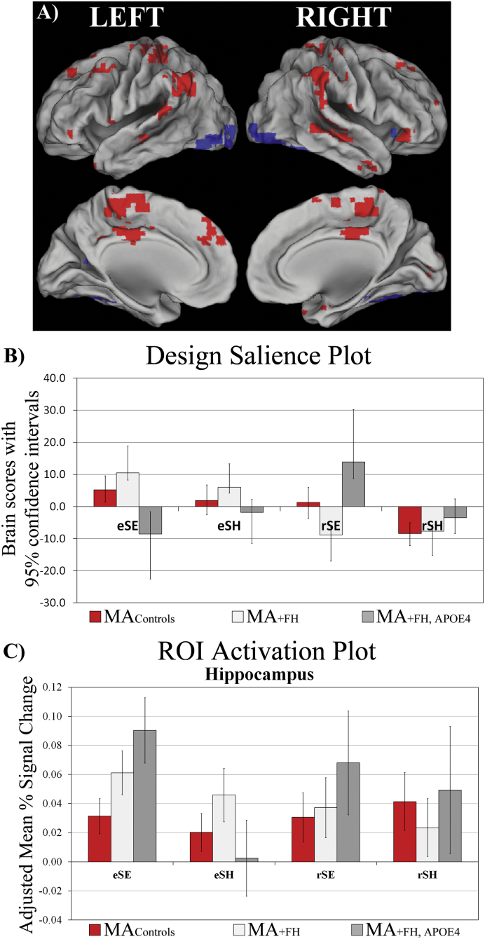
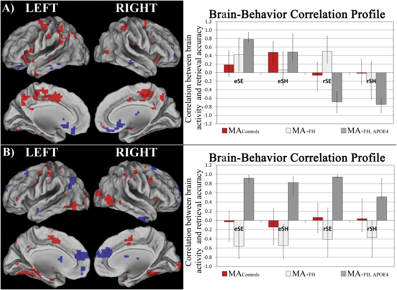
Episodic memory impairment is a consistent, pronounced deficit in pre-clinical stages of late-onset Alzheimer's disease (AD). Individuals with risk factors for AD exhibit altered brain function several decades prior to the onset of AD-related symptoms. In the current event-related fMRI study of spatial context memory we tested the hypothesis that middle-aged adults (MA; 40-58 yrs) with a family history of late onset AD (MA+ FH), or a combined + FH and apolipoprotein E ε4 allele risk factors for AD (MA+ FH + APOE4), will exhibit differences in encoding and retrieval-related brain activity, compared to - FH - APOE4 MA controls. We also hypothesized that the two at-risk MA groups will exhibit distinct patterns of correlation between brain activity and memory performance, compared to controls. To test these hypotheses we conducted multivariate task, and behavior, partial least squares analysis of fMRI data obtained during successful context encoding and retrieval. Our results indicate that even though there were no significant group differences in context memory performance, there were significant differences in brain activity and brain-behavior correlations involving the hippocampus, inferior parietal cortex, cingulate, and precuneus cortex in MA with AD risk factors, compared to controls. In addition, we observed that brain activity and brain-behavior correlations in anterior-medial PFC and in ventral visual cortex differentiated the two MA risk groups from each other, and from MAcontrols. Our results indicate that functional differences in episodic memory-related regions are present by early midlife in adults with + FH and + APOE-4 risk factors for late onset AD, compared to middle-aged controls.


Figures

Fig. 1
Fig. 1
T-PLS LV1 and LV2 result. A) The singular image and design salience plot for T-PLS LV1. The singular image is thresholded at a bootstrap ratio of ± 3.5, p < 0.001. Red brain regions reflect positive brain saliences and blue regions reflect negative brain saliences. Activations are presented on template images of the lateral and medial surfaces of the left and right hemispheres of the brain using Caret software (http://brainvis.wustl.edu/wiki/index.php/Caret:Download). The design salience plots represent the brain scores with 95% confidence intervals (y-axis) for each group for each task-type (x-axis). eSE = encoding, easy spatial context memory tasks; eSH = encoding, hard spatial context memory tasks; rSE = retrieval, easy spatial context memory tasks; rSH = retrieval, hard spatial context memory tasks. The design salience plot for T-PLS LV1 indicates this LV identified brain regions that were differentially activated during successful spatial context retrieval (positive saliences) vs. encoding (negative saliences). B) The singular image and design salience plot for T-PLS LV2. The singular image is thresholded at a bootstrap ratio of ± 3.5, p < 0.001. This LV identified brain regions that were differentially activated during hard spatial encoding (eSH; positive saliences) vs. easy spatial encoding (eSE; negative saliences).
Fig. 2
Fig. 2
T-PLS LV3 and right hippocampal activation plot. A) The singular image and B) design salience plot for T-PLS LV3. The singular image is thresholded at a bootstrap ratio of ± 3.5, p < 0.001. Red brain regions reflect positive brain saliences and blue regions reflect negative brain saliences. The design salience plot for T-PLS LV3 indicates this LV identified a group ∗ task ∗ phase interaction. Positive salience brain regions were more active during encoding > retrieval in MA+ FH and MAcontrols; and more activity during easy spatial context retrieval > easy spatial context encoding in MA+ FH + APOE4. Negative salience brain regions exhibited the inverse pattern of associations. C) ROI activation plot for right hippocampus from T-PLS LV2.
Fig. 3
Fig. 3
B-PLS results. A) The singular image at a bootstrap ratio threshold = ± 3.5, p < 0.001 and the brain-behavior correlation profile with 95% confidence intervals for B-PLS LV1. In the singular image red brain regions reflect positive brain saliences and blue regions reflect negative brain saliences. Activations are presented on template images of the lateral and medial surfaces of the left and right hemispheres of the brain using Caret software. The correlation profile indicates that encoding activity in positive salience regions was positively correlated with subsequent retrieval for both task in MA+ FH + APOE4 and MAcontrols; and retrieval activity in these same regions was negative correlated with retrieval accuracy on both tasks in MA+ FH + APOE4. In MA+ FH encoding and retrieval activity in positive salience regions during SE tasks was correlated with better performance on this task. Negative salience regions exhibited the inverse pattern of brain-behavior correlations. B) The singular image at a bootstrap ratio threshold = ± 3.5, p < 0.001 and the brain-behavior correlation profile with 95% confidence intervals for B-PLS LV2. The correlation profile indicates that increased encoding and retrieval activity in positive salience regions was positively correlated with memory performance on both tasks in MA+ FH + APOE4 subjects, and negatively correlated with memory performance on both tasks in MA+ FH. Negative salience regions exhibited the inverse pattern of brain-behavior correlations.

References

    1. Agena-Bioscience . In: The Science of SNP Genotyping. Bioscience Agena, Genome Quebec A.B., editors. Agena Bioscience; San Diego, CA, USA: 2015.
    1. Alexander G.E. Gray matter network associated with risk for Alzheimer's disease in young to middle-aged adults. Neurobiol. Aging. 2012;33(12):2723–2732. - PMC - PubMed
    1. Ankudowich E., Pasvanis S., Rajah M.N. Changes in the modulation of brain activity during context encoding vs. context retrieval across the adult lifespan. NeuroImage. 2016;139:103–113. - PubMed
    1. Backman L. Brain regions associated with episodic retrieval in normal aging and Alzheimer's disease. Neurology. 1999;52(9):1861–1870. - PubMed
    1. Backman L. Multiple cognitive deficits during the transition to Alzheimer's disease. J. Intern. Med. 2004;256(3):195–204. - PubMed

Publication types

MeSH terms

Grants and funding