

Preclinical imaging of iridocorneal angle and fundus using a modified integrated flexible handheld probe
- PMID: 28413809
- PMCID: PMC5386004
- DOI: 10.1117/1.JMI.4.2.026001
Preclinical imaging of iridocorneal angle and fundus using a modified integrated flexible handheld probe
Abstract
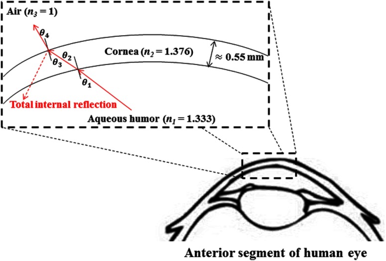
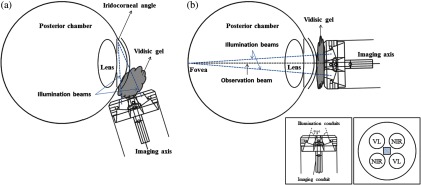
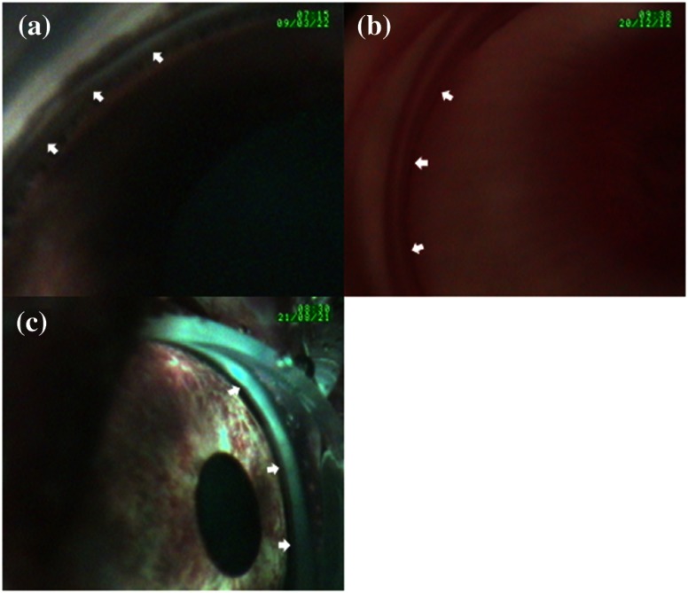
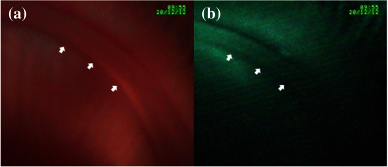
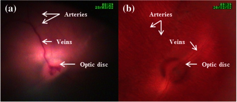
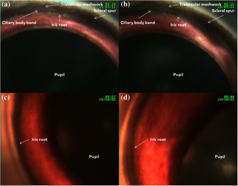
A flexible handheld imaging probe consisting of a [Formula: see text] charge-coupled device camera, light-emitting diode light sources, and near-infrared laser source is designed and developed. The imaging probe is designed with specifications to capture the iridocorneal angle images and posterior segment images. Light propagation from the anterior chamber of the eye to the exterior is considered analytically using Snell's law. Imaging of the iridocorneal angle region and fundus is performed on ex vivo porcine samples and subsequently on small laboratory animals, such as the New Zealand white rabbit and nonhuman primate, in vivo. The integrated flexible handheld probe demonstrates high repeatability in iridocorneal angle and fundus documentation. The proposed concept and methodology are expected to find potential application in the diagnosis, prognosis, and management of glaucoma.
Keywords: fundus imaging; imaging systems; iridocorneal angle imaging; medical imaging; ophthalmology.
Figures
Similar articles
-
Integrated flexible handheld probe for imaging and evaluation of iridocorneal angle.J Biomed Opt. 2015 Jan;20(1):016014. doi: 10.1117/1.JBO.20.1.016014. J Biomed Opt. 2015. PMID: 25594626
-
Note: A gel based imaging technique of the iridocorneal angle for evaluation of angle-closure glaucoma.Rev Sci Instrum. 2014 Jun;85(6):066105. doi: 10.1063/1.4882335. Rev Sci Instrum. 2014. PMID: 24985869
-
Noninvasive and Noncontact Sequential Imaging of the Iridocorneal Angle and the Cornea of the Eye.Transl Vis Sci Technol. 2020 Apr 9;9(5):1. doi: 10.1167/tvst.9.5.1. eCollection 2020 Apr. Transl Vis Sci Technol. 2020. PMID: 32821473 Free PMC article.
-
[Diacaustic examination of the iridocorneal angle : Video article].Ophthalmologe. 2018 Jul;115(7):606-612. doi: 10.1007/s00347-018-0741-x. Ophthalmologe. 2018. PMID: 29948150 Review. German.
-
[A new approach for studying the retinal and choroidal circulation].Nippon Ganka Gakkai Zasshi. 2004 Dec;108(12):836-61; discussion 862. Nippon Ganka Gakkai Zasshi. 2004. PMID: 15656089 Review. Japanese.
References
LinkOut - more resources
Full Text Sources
Other Literature Sources
