Review
. 2018 Jan;1860(1):83-90.
doi: 10.1016/j.bbamem.2017.04.008. Epub 2017 Apr 13.

Spatio-temporal regulation of connexin43 phosphorylation and gap junction dynamics

Affiliations
Review

Spatio-temporal regulation of connexin43 phosphorylation and gap junction dynamics

Joell L Solan et al. Biochim Biophys Acta Biomembr. 2018 Jan.

Abstract

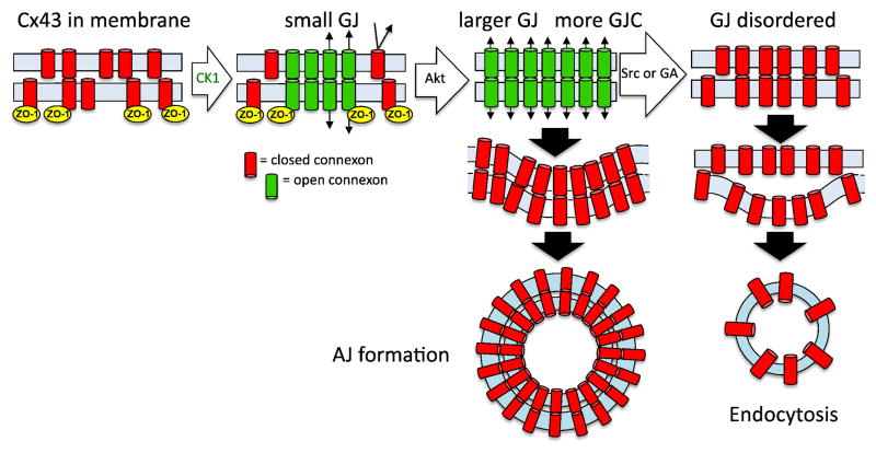
Gap junctions are specialized membrane domains containing tens to thousands of intercellular channels. These channels permit exchange of small molecules (<1000Da) including ions, amino acids, nucleotides, metabolites and secondary messengers (e.g., calcium, glucose, cAMP, cGMP, IP3) between cells. The common reductionist view of these structures is that they are composed entirely of integral membrane proteins encoded by the 21 member connexin human gene family. However, it is clear that the normal physiological function of this structure requires interaction and regulation by a variety of proteins, especially kinases. Phosphorylation is capable of directly modulating connexin channel function but the most dramatic effects on gap junction activity occur via the organization of the gap junction structures themselves. This is a direct result of the short half-life of the primary gap junction protein, connexin, which requires them to be constantly assembled, remodeled and turned over. The biological consequences of this remodeling are well illustrated during cardiac ischemia, a process wherein gap junctions are disassembled and remodeled resulting in arrhythmia and ultimately heart failure. This article is part of a Special Issue entitled: Gap Junction Proteins edited by Jean Claude Herve.

Keywords: Connexin43; Gap junction; Kinase; Phosphorylation.


Figures

Figure 1
Figure 1
A model of gap junction assembly and disassembly. Under homeostatic conditions, connexons aggregate into gap junctions via interaction with ZO-1 and CK1. Acute disassembly can be induced by a variety of stimuli that initially lead to Akt-mediated phosphorylation on Cx43, which inhibits ZO-1 interaction, and permits gap junction growth. Depending on stimuli and phosphorylation status of Cx43, internalization can occur via annular junction (AJ) formation or through disruption of gap junction organization and channel coupling then subsequent endocytosis.
Figure 2
Figure 2
Rapid changes in Cx43 phosphorylation occur in response to compounds that induce gap junction disassembly. Immunoblot analysis of NRK lysates with antibody to total Cx43 shows loss of the P0 form of Cx43 within 5 minutes of treatment with EGF 20ng/ml or TPA 50nM (Sigma). Linescans of the band densities are shown on the right. Antibodies and methods used are as previously described [104].
Figure 3
Figure 3
Spatiotemporal changes in gap junction size and phosphorylation during induced disassembly. NRK cells were fixed and stained with a mouse antibody to total Cx43 (green) and a rabbit antibody to Cx43 phosphorylated on S373 (red). Gap junctions in control cells range in size and show little signal for pS373. After 5 minutes of TPA treatment (100nM) gap junction size is increased and Cx43 is phosphorylated on S373 (yellow). After 30 minutes of treatment gap junctions are diminished, disorganized and in various states of internalization. Antibodies and methods used were as described in [53]. (bar=25μm).
Figure 4
Figure 4
Phosphorylation of Cx43 on S262, S279/282 accumulates upon treatment of NRK cells with EGF or TPA, while S368 is only phosphorylated in response to TPA. NRK cell lysates taken at various times after treatment were analyzed by immunoblot with antibodies to total Cx43 and rabbit antibodies to S262 (Santa Cruz Biotechnology), 279/282 [76] and S368 (R&D systems). Antibodies and methods used were as described in [76]. The position of the 50kDa marker is indicated on the right side.
Figure 5
Figure 5
Phosphorylation on S262 correlates with activation of ERK. NRK cells were treated with EGF and TPA for 30 minutes in the presence of the PKC inhibitor bisindomaleimide (BIM) or the ERK inhibitor PD98059 (PD) (Calbiochem). A. Cell lysates were analyzed by immunoblot with antibodies to S262, S279/282, S368 and ERK phosphorylated on Thr202/Tyr204 (Cell Signaling Technologies). The positions of the 50kDa and 36kDa (for the 4-Cx43 blots) are shown on the right. B. Graph shows quantification of Cx43 phosphorylation levels (n=3) and the ratio of pERK/total ERK is shown below. Note the scale on the left refers to S279/282 and S368 relative phosphorylation levels and the one on the right, which is non-linear, to S262. Linear regression analysis shows the phosphorylation on S262 is correlated to ERK phosphorylation with an R2=0.812.

References

    1. Severs NJ. The cardiac gap junction and intercalated disc. Int J Cardiol. 1990;26(2):137–73. - PubMed
    1. Muller DJ, et al. Conformational changes in surface structures of isolated connexin 26 gap junctions. EMBO J. 2002;21(14):3598–607. - PMC - PubMed
    1. Goodenough DA, Goliger JA, Paul DL. Connexins, connexons, and intercellular communication. Ann Rev Biochem. 1996;65:475–502. - PubMed
    1. Bruzzone R, White TW, Paul DL. Connections with connexins: The molecular basis of direct intercellular signaling. Eur J Biochem. 1996;238:1–27. - PubMed
    1. Willecke K, et al. Structural and functional diversity of connexin genes in the mouse and human genome. Biol Chem. 2002;383:725–737. - PubMed

Publication types