. 2017 Aug 17;36(33):4810-4816.
doi: 10.1038/onc.2017.106. Epub 2017 Apr 17.

Transforming activity and therapeutic targeting of C-terminal-binding protein 2 in Apc-mutated neoplasia

Affiliations

Transforming activity and therapeutic targeting of C-terminal-binding protein 2 in Apc-mutated neoplasia

E T Sumner et al. Oncogene. .

Abstract

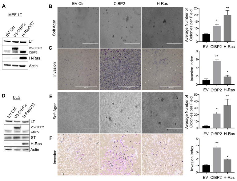
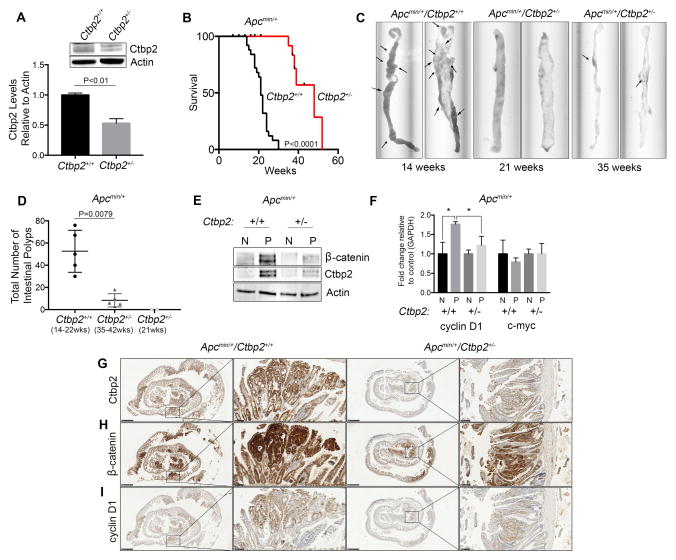
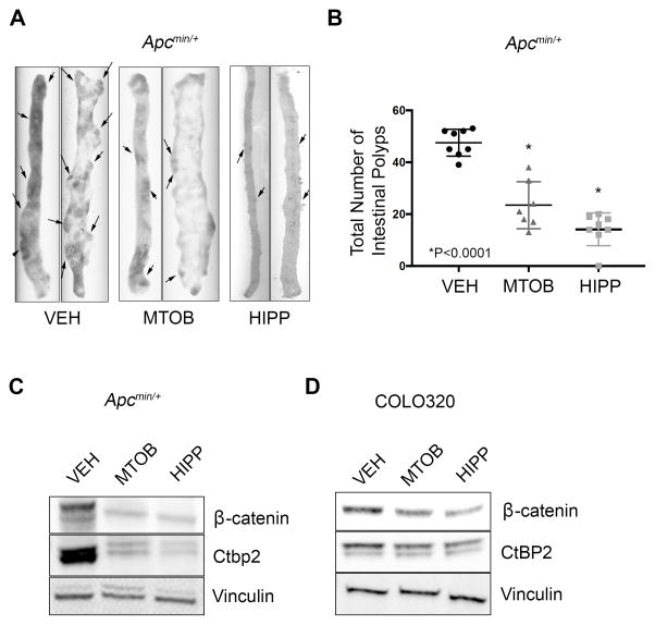
Overexpression of the transcriptional coregulators C-terminal binding proteins 1 and 2 (CtBP1 and 2) occurs in many human solid tumors and is associated with poor prognosis. CtBP modulates oncogenic gene expression programs and is an emerging drug target, but its oncogenic role is unclear. Consistent with this oncogenic potential, exogenous CtBP2 transformed primary mouse and human cells to anchorage independence similarly to mutant H-Ras. To investigate CtBP's contribution to in vivo tumorigenesis, Apcmin/+ mice, which succumb to massive intestinal polyposis, were bred to Ctbp2+/- mice. CtBP interacts with adenomatous polyposis coli (APC) protein, and is stabilized in both APC-mutated human colon cancers and Apcmin/+ intestinal polyps. Ctbp2 heterozygosity increased the median survival of Apcmin/+ mice from 21 to 48 weeks, and reduced polyp formation by 90%, with Ctbp2+/- polyps exhibiting reduced levels of β-catenin and its oncogenic transcriptional target, cyclin D1. CtBP's potential as a therapeutic target was studied by treating Apcmin/+ mice with the CtBP small-molecule inhibitors 4-methylthio-2-oxobutyric acid and 2-hydroxy-imino phenylpyruvic acid, both of which reduced polyposis by more than half compared with vehicle treatment. Phenocopying Ctbp2 deletion, both Ctbp inhibitors caused substantial decreases in the protein level of Ctbp2, as well its oncogenic partner β-catenin, and the effects of the inhibitors on CtBP and β-catenin levels could be modeled in an APC-mutated human colon cancer cell line. CtBP2 is thus a druggable transforming oncoprotein critical for the evolution of neoplasia driven by Apc mutation.


Conflict of interest statement

Conflict of Interest

The authors have declared that no conflict of interest exists.

Figures

Figure 1
Figure 1. CtBP2 transforms primary mouse and human cells
(A–C) CtBP2 cooperates with SV40 Large T-antigen to induce transformation of primary MEFs. (A) Immunoblot with indicated antibodies of LT-expressing MEFs infected with indicated retroviruses. Endogenous CtBP2 and V5-CtBP2 bands are indicated. (B) Soft-agar colony formation assay of LT-expressing MEFs infected with the indicated retroviruses (*p<0.05). (C) Invasion assay of LT-expressing MEFs infected with indicated retroviruses (*p<0.01). (D–F) CtBP2 cooperates with both SV40 Large T and Small T-antigens to transform human fibroblasts. (D) Immunoblot with indicated antibodies of LT/ST-expressing BJ-hTERT cells infected with indicated retroviruses. Endogenous CtBP2 and V5-CtBP2 bands are indicated. (E) Soft-agar colony formation assay of LT/ST-expressing BJ-hTERT cells infected with the indicated retroviruses (*p<0.05) (F) Invasion assay of LT/ST-expressing BJ-hTERT cells infected with indicated retroviruses (*p<0.01). Scale bars represent 400um. Statistical analyses were performed by t-test. Error bars represent ±SD of three independent experiments performed in triplicate. Methods: BJ-hTERT-Blast cells (tested and confirmed mycoplasma free) were kindly provided by L. Litovchick and were maintained in EMEM (GIBCO) supplemented with 10% FBS and incubated at 37ºC in a humidified, 5% CO2 environment. Primary MEFs were obtained from mouse embryos at age E10.5. After harvest, cells were plated onto gelatin-coated plates and left to attach for 24 hours before use in assays. MEFs were maintained in DMEM supplemented with 10% FBS, 5% MEM non-essential amino acids (Life Technologies), and 5% L-glutamine (Life Technologies). MEF cells infected with CtBP2 or H-RasV12-expressing retroviruses were selected for one week using 1μg/ml puromycin. MEF cells infected with LT (cDNA)-expressing retrovirus were selected for two weeks using 200 μg/ml zeocin. BJ-hTERT cells were serially infected with specified plasmids and selected with puromycin (CtBP2, H-Ras) and/or zeocin (LT/ST genomic construct). For invasion assays, cells were plated into 6-well Matrigel-coated invasion chambers (Corning). After 24 hours, chambers were washed in PBS, fixed in methanol for 20 minutes and stained in Giemsa solution overnight. For soft agar assays, cells expressing specified plasmids were mixed into 0.3% noble agar and poured onto 60 mm plates containing a solidified bottom layer of 0.6% noble agar. Plates were left to form colonies for 21 days. For western blotting, cells were washed in cold PBS and lysed in RIPA buffer (150mM NaCl, 50mM Tris HCL, pH 8.0, 1.7% NP-40, 0.17% sodium dodecyl sulfate (SDS), 0.5% Na-deoxycholate monohydrate, 5mM EDTA) containing 1 tablet of complete Mini Protease Inhibitor Cocktail/10ml (Roche). Lysates were cleared of insoluble material by centrifugation at 15,000 RPM. Proteins (30 μg) were loaded onto a Bis-Tris 4–12% gel containing NuPAGE MES buffer. pBABE-zeo-LargeTgenomic (expressing LT and ST), pBABE-zeo-LargeTcDNA, and pBABE-puro-H-Ras were obtained from Addgene. CtBP2 cDNA was cloned into pBABE-puro-V5 to produce pBABE-puro-CtBP2V5. Pc-CtBP2V5 has been previously described . Antibodies used were as follows: anti-CtBP2 E-16 (Santa Cruz Biotechnologies, Santa Cruz, CA); anti-SV40-LT Pab 101 (Santa Cruz); anti-SV40-ST Pab 419 (Santa Cruz); anti-Actin (Sigma Aldrich); anti-Ras C-20 (Santa Cruz).
Figure 2
Figure 2. Ctbp2 haploinsuffciency in Apcmin/+ mice reduces polyp formation and increases survival
(A) Intestinal tissue from Ctbp2+/+ and Ctbp2+/− mice was immunoblotted using anti-Ctbp2 antibody and Ctbp2 abundance determined by densitometry. Representative immunoblot shown of 3 independent biologic replicates. (B) Overall survival of Apcmin/+/Ctbp2+/+ (n=25) vs Apcmin/+/Ctbp2+/− mice (n=20). Median survival for Ctbp+/+ mice was 21 weeks while median survival for Ctbp2+/− was 39 weeks (log-rank test, p<0.0001). (C) Representative small intestines from Apcmin/+/Ctbp2+/+ or Apcmin/+/Ctbp2+/−mice sacrificed at indicated times (arrows represent polyps). (D) Box plot comparing total intestinal polyp numbers from Apcmin/+/Ctbp2+/+ or Apcmin/+/Ctbp2+/−mice at the indicated ages. Median intestinal polyp numbers found in mice at time of death, sacrifice at humane endpoint, or censoring were Ctbp2+/+ = 56 and Ctbp2+/− = 5 (n=5 per group). *** Apcmin/+/Ctbp2+/− mice at 21 weeks (n=2) had no visible polyps. Statistical analysis employed the Mann-Whitney test. (E) Immunoblot (representative of 3 biologic replicates) demonstrating Ctbp2 and β-catenin protein levels from normal (N) and polyp (P) intestinal tissue obtained from Apcmin/+/Ctbp2+/+ or Apcmin/+/Ctbp2+/−mice obtained at 21 weeks of age and 37 weeks of age. (F) Q-PCR analysis on mRNA derived from normal (N) and polyp (P) tissue from both groups of mice, probing for mouse c-myc and cyclin-D1 (t-test, *p<0.05). Error bars for Q-PCR represent ± SEM of two independent experiments performed in triplicate. (G–I) Immunohistochemical staining of intestinal tissue for (G) Ctbp2, (H) β-catenin, (I) cyclin D1. Representative sections shown from polyps analyzed from at least 3 different animals. Scale bars represent 1mm and 100μm. Methods: Apcmin/+ mice (C57BL/6J, Jackson Labs) were crossed with Ctbp2+/− mice (B6;129S4 X C57BL/6J backcrossed > 6 generations, Jackson Labs) to generate mice harboring the min mutation and either WT Ctbp2 (Apcmin/+/Ctbp2+/+) or hemizygous Ctbp2 (Apcmin/+/Ctbp2+/−). Sample sizes for each cohort were estimated to ensure 80% power. All mouse experiments were performed with approval of the VCU IACUC. To confirm desired genotypes of study mice, the following primers were used for endpoint PCR: Apcmin/+ F: GGGAAGTTTAGACAGTTCTCGT, R: TGTTGGATGGTAAGCACTGAG, Mut: AGACAGAAGTTAGGAGAGAGAGC, WT: AGACAGAAGTTTGGAGAGAGAGC. Ctbp2+/− Internal Con F: 5′-CAAATGTTGCTTGTCTGGTG-3′, Internal Con R: 5′-GTCAGTCGAGTGCACAGTTT-3′, Mut F: 5′-CCAGTGGGGATCGACGGTATC-3′, Mut R: CACTCCAACGCAGCACCATC. Survival curves were generated from mice that were monitored daily and euthanized when moribund. In the case of Apcmin/+/Ctbp2+/− mice in the survival study, six mice were sacrificed before humane endpoint (while still healthy, counted as censored) to obtain intestinal tissues (2 at 21 weeks, 3 at 37 weeks, and 1 at 38 weeks). Ctbp2+/+ intestinal tissues, used for polyp number, protein, mRNA, and histopathological analysis, were obtained at time of death (or humane endpoint). Whole intestines were surgically removed, perfused with PBS, and analyzed for polyp number. For H&E staining, intestines were cut into 4–5cm strips and perfused with PBS followed by 10% formalin solution. Once clean, strips were cut longitudinally on blotting paper and Swiss-roll preparations were stained with H&E. For protein and mRNA analysis, polyps (and adjacent normal tissue) were cut into roughly 1 cm3 pieces and ground by mortar and pestle followed by resuspension in either RIPA supplemented with protease inhibitors for protein analysis, or RNAeasy buffer (Qiagen) to obtain mRNA. To examine changes of protein level and localization of Cyclin D, CtBP2, and β-catenin, immunohistochemical analysis was performed using mouse anti-Cyclin D (72-13G, Santa Cruz), β-catenin and CtBP2 (Clone 14 and 16, respectively; BD BioSciences); each were used at a 1:50 dilution. The paraffin-embedded tissue was de-paraffinized in xylene and rehydrated through graded ethanol, followed by antigen retrieval using 10 mM sodium citrate buffer (pH 6.0) for 15 min. The slides were allowed to cool to room temperature for 30 min. After the slides were washed, exogenous peroxidase activity was blocked by incubating the slides in 3% peroxidase for 20 min. The slides were then washed, blocked in 5% NGS-PBS for 60 min at room temperature, and incubated overnight at 4°C in primary antibody diluted in 5% NGS-PBS. Following primary antibody incubation and washing with PBS, the slides were incubated in secondary HRP anti-mouse IgG reagent (Dako Agilent Technologies) at room temperature for 1 hour, washed with PBS and then detected using EnVision+ kit (Dako Agilent Technologies). Counterstaining with hematoxylin was performed and the slides were dehydrated through graded ethanol and then xylene and mounted with Entelen (Electron Microscopy, Inc.). Total cell RNA was purified using RNeasy (Qiagen) according to manufacturer’s suggested protocol. cDNA was generated using Affinity Script RT (Agilent Technologies). One μL cDNA was amplified in triplicate using SYBRgreen and the specified primers (see Supplemental Information). Q-PCR primers: c-myc (F5′-AACAGGAACTATGACCTCG-3′, R5′-AGCAGCTCGAATTTCTTC-3′) cyclin-d1 (F5′-CAGACGTTCAGAACCAGATTC-3′, R5′-CCCTCCAATAGCAGCGAAAAC-3′). GAPDH (F5′-AAGGTCATCCCAGAGCTGAA-3′, R5′-CTGCTTCACCACCTTCTT-3′).
Figure 3
Figure 3. Effect of the Ctbp2 inhibitors MTOB and HIPP on Apcmin/+ polyposis
(A) Representative small intestine tissue from Apcmin/+ mice treated with VEH-control, MTOB, or HIPP. (B) Box plot comparing total intestinal polyp numbers found in mice at time of sacrifice. Median numbers were VEH = 45, MTOB = 21, and HIPP = 15 (n=7 per group; *p<0.0001, Mann-Whitney test). (C) Immunoblot of Ctbp2 and β-catenin protein levels in polyps recovered from Apcmin/+ mice treated with VEH-control, MTOB, or HIPP. (D) Immunoblot demonstrating CtBP2 and β-catenin levels from COLO320 cells treated with VEH-control, MOTB, or HIPP. Immunoblots in C and D are representative of 3 biologic replicates. Methods: For MTOB/HIPP studies, Apcmin/+ mice received IP injections (3X weekly for 8 weeks) of VEH-control (0.2M NaHO3 solution in water), 750mg/kg MTOB (dissolved in 0.2M NaHO3/water solution; Sigma-Aldrich), or 250mg/kg HIPP (dissolved in 0.2M NaHO3/water solution). Sample sizes for each cohort were estimated to ensure 80% power. Mice with the correct genotype were randomly assigned to the vehicle or drug group. COLO320 cells were obtained from ATCC (confirmed mycoplasma free) and maintained in RPMI-1640 supplemented with 10% FBS and incubated at 37ºC in a humidified, 5% CO2 environment.

References

    1. Turner J, Crossley M. The CtBP family: enigmatic and enzymatic transcriptional co-repressors. Bio Essays News Rev Mol Cell Dev Biol. 2001;23:683–690. - PubMed
    1. Chinnadurai G. The transcriptional corepressor CtBP: a foe of multiple tumor suppressors. Cancer Res. 2009;69:731–734. - PMC - PubMed
    1. Di L-J, Byun JS, Wong MM, Wakano C, Taylor T, Bilke S, et al. Genome-wide profiles of CtBP link metabolism with genome stability and epithelial reprogramming in breast cancer. Nat Commun. 2013;4:1449. - PMC - PubMed
    1. Zhang C, Gao C, Xu Y, Zhang Z. CtBP2 could promote prostate cancer cell proliferation through c-Myc signaling. Gene. 2014;546:73–79. - PubMed
    1. Barroilhet L, Yang J, Hasselblatt K, Paranal RM, Ng S-K, Rauh-Hain JA, et al. C-terminal binding protein-2 regulates response of epithelial ovarian cancer cells to histone deacetylase inhibitors. Oncogene. 2013;32:3896–3903. - PubMed

MeSH terms