. 2017 May;23(5):638-643.
doi: 10.1038/nm.4319. Epub 2017 Apr 17.

HIV persistence in tissue macrophages of humanized myeloid-only mice during antiretroviral therapy

Affiliations

HIV persistence in tissue macrophages of humanized myeloid-only mice during antiretroviral therapy

Jenna B Honeycutt et al. Nat Med. 2017 May.

Abstract

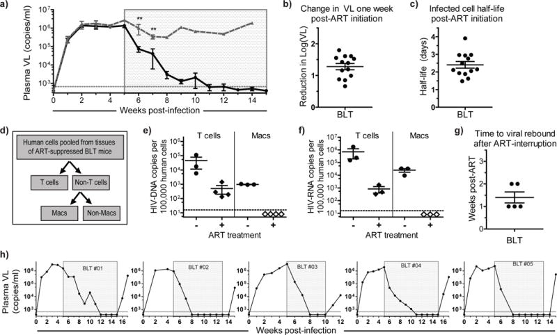
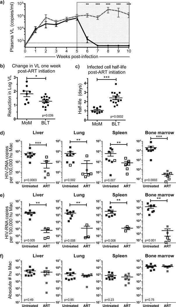
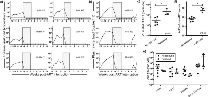
Despite years of fully suppressive antiretroviral therapy (ART), HIV persists in its hosts and is never eradicated. One major barrier to eradication is that the virus infects multiple cell types that may individually contribute to HIV persistence. Tissue macrophages are critical contributors to HIV pathogenesis; however, their specific role in HIV persistence during long-term suppressive ART has not been established. Using humanized myeloid-only mice (MoM), we demonstrate that HIV infection of tissue macrophages is rapidly suppressed by ART, as reflected by a rapid drop in plasma viral load and a dramatic decrease in the levels of cell-associated viral RNA and DNA. No viral rebound was observed in the plasma of 67% of the ART-treated animals at 7 weeks after ART interruption, and no replication-competent virus was rescued from the tissue macrophages obtained from these animals. In contrast, in a subset of animals (∼33%), a delayed viral rebound was observed that is consistent with the establishment of persistent infection in tissue macrophages. These observations represent the first direct evidence, to our knowledge, of HIV persistence in tissue macrophages in vivo.


Conflict of interest statement

The authors have no competing interests as defined by Springer Nature, or other interests that might be perceived to influence the results and/or discussion reported in this paper.

Figures

Figure 1
Figure 1. Viral suppression, persistence and structured ART-interruption induced rebound in BLT mice
a) Plasma viral load (VL) was monitored over time in HIV-infected ART-treated (n=13, solid black line) and untreated (n=5, dashed gray line) BLT mice. Each point represents the mean ± s.e.m. The time of exposure to ART for the treated animals is indicated with a shaded gray box. A Mann-Whitney test was used to compare the plasma VL of ART-treated and untreated mice at one and two weeks post-ART initiation (p=0.0049 and p=0.0080, respectively). b) The reduction in log (base 10) plasma VL one week after ART-initiation was calculated for each treated BLT mouse (n=13). c) The half-life of productively infected cells was estimated from the change in viral load during ART (n=13). d) Schematic for the magnetic sorting and purification of human T cells and macrophages from the tissues obtained from infected animals. Human T cells (CD3+) and non-T cells were separated from pooled tissue cells of individual ART-suppressed BLT mice (n=4). Macrophages (Macs) were then isolated from the non-T cell fraction. Analysis of (e) HIV-DNA and (f) HIV-RNA levels were performed by real time PCR using purified cells isolated from ART-suppressed (n=4) and untreated (n=3) BLT mice. HIV-DNA/RNA levels are normalized and reported per 100,000 human cells. Samples with values below the level of detection are indicated by open diamonds in e & f and are shown at the average lower limit of detection. g) Time to viral rebound after ART interruption in BLT mice (n=5). h) Viral rebound was observed 1–2 weeks after ART-interruption in all BLT mice. For b, c, e, f & g, the horizontal lines represent mean ± s.e.m.
Figure 2
Figure 2. ART rapidly suppresses viral replication in MoM
a) Longitudinal analysis of the plasma VL in HIV-infected ART-treated (n=8, solid black line) and untreated (n=6, dashed gray line) MoM. Each point represents the mean ± s.e.m.. A Mann-Whitney test was used to compare the plasma VL of treated and untreated mice at 1–5 weeks post-ART initiation (p=0.0019, p=0.0008, p=0.0008, p=0.0011 and p=0.0017, respectively). b) The reduction in log (base 10) plasma VL one week after ART-initiation was calculated for each treated MoM (n=8). c) The half-life of productively infected cells was estimated from the change in viral load during ART (n=8). A Mann-Whitney test was used to compare MoM and BLT mice in b & c. Cell-associated (d) HIV-DNA and (e) HIV-RNA levels were measured in the liver, lung, spleen and bone marrow of ART-treated (n=6, gray squares) and untreated (n=8, black squares) MoM. Undetectable samples are indicated by an empty black box shown at the limit of detection for that sample (dependent on the number of cells available for analysis). Viral DNA and RNA levels were normalized per 100,000 human macrophages and compared between treated and untreated mice. A log-rank test was used to account for censoring due to the limits of detection in d & e. f) There was no difference in the total numbers of human macrophages present in the tissues of ART-treated or untreated MoM (all p>0.05, Mann-Whitney test). In b–f, horizontal lines represent mean ± s.e.m.
Figure 3
Figure 3. HIV persistence in tissue macrophages during ART
Viral rebound was absent in most infected MoM after structured ART-interruption [(a) n=6 No rebound, (b) n=3 Rebound]. c) Higher plasma VL at the start of treatment and (d) higher total viral burden [as demonstrated by area under the curve (AUC) analysis of pre-ART viremia] was associated with viral rebound after ART-interruption. e) The total numbers of human macrophages in the tissues of MoM were similar between mice where viral rebound was absent (a) or observed (b) (p>0.05). Mann-Whitney tests were used to compare mice in c–e. For c–e, the horizontal lines represent mean ± s.e.m.

Comment in

References

    1. Kumar A, Herbein G. The macrophage: a therapeutic target in HIV-1 infection. Mol Cell Ther. 2014;2:10. - PMC - PubMed
    1. Koppensteiner H, Brack-Werner R, Schindler M. Macrophages and their relevance in Human Immunodeficiency Virus Type I infection. Retrovirology. 2012;9:82. - PMC - PubMed
    1. Campbell JH, Hearps AC, Martin GE, Williams KC, Crowe SM. The importance of monocytes and macrophages in HIV pathogenesis, treatment, and cure. AIDS. 2014;28:2175–2187. - PMC - PubMed
    1. Sattentau QJ, Stevenson M. Macrophages and HIV-1: An Unhealthy Constellation. Cell Host Microbe. 2016;19:304–310. - PMC - PubMed
    1. Collman RG, Perno CF, Crowe SM, Stevenson M, Montaner LJ. HIV and cells of macrophage/dendritic lineage and other non-T cell reservoirs: new answers yield new questions. J Leukoc Biol. 2003;74:631–634. - PubMed

MeSH terms