. 2017 Apr 17;12(4):e0175835.
doi: 10.1371/journal.pone.0175835. eCollection 2017.

Sustained low peritoneal effluent CCL18 levels are associated with preservation of peritoneal membrane function in peritoneal dialysis

Affiliations

Sustained low peritoneal effluent CCL18 levels are associated with preservation of peritoneal membrane function in peritoneal dialysis

Marta Ossorio et al. PLoS One. .

Abstract

Peritoneal membrane failure (PMF) and, ultimately, encapsulating peritoneal sclerosis (EPS) are the most serious peritoneal dialysis (PD) complications. Combining clinical and peritoneal transport data with the measurement of molecular biomarkers, such as the chemokine CCL18, would improve the complex diagnosis and management of PMF. We measured CCL18 levels in 43 patients' effluent and serum at baseline and after 1, 2, and 3 years of PD treatment by retrospective longitudinal study, and evaluated their association with PMF/EPS development and peritoneal risk factors. To confirm the trends observed in the longitudinal study, a cross-sectional study was performed on 61 isolated samples from long-term (more than 3 years) patients treated with PD. We observed that the patients with no membrane dysfunction showed sustained low CCL18 levels in peritoneal effluent over time. An increase in CCL18 levels at any time was predictive of PMF development (final CCL18 increase over baseline, p = .014; and maximum CCL18 increase, p = .039). At year 3 of PD, CCL18 values in effluent under 3.15 ng/ml showed an 89.5% negative predictive value, and higher levels were associated with later PMF (odds ratio 4.3; 95% CI 0.90-20.89; p = .067). Moreover, CCL18 levels in effluent at year 3 of PD were independently associated with a risk of PMF development, adjusted for the classical (water and creatinine) peritoneal transport parameters. These trends were confirmed in a cross-sectional study of 61 long-term patients treated with PD. In conclusion, our study shows the diagnostic capacity of chemokine CCL18 levels in peritoneal effluent to predict PMF and suggests CCL18 as a new marker and mediator of this serious condition as well as a new potential therapeutic target.


Conflict of interest statement

Competing Interests: The authors have declared that no competing interests exist.

Figures

Fig 1
Fig 1. Low effluent CCL18 concentrations over time are found in patients with no peritoneal membrane failure.
(A) Time course of effluent CCL18 concentrations define two groups of patients on PD. Eleven patients were included in Group 1, and 32 patients were included in group 2. Mean ± SD values are shown. Higher CCL18 effluent values were found in group 2 at all time points **p < .005; ***p < .001 (Mann-Whitney U test). (B) Kaplan-Meier analysis of PMF development in the patients included in group 1 and group 2. Patients with no sample at a given time point were censored. Log rank test p = .076. (C) Mean Cr D/P and peritoneal protein losses in patients included in Group 1 and Group 2 evaluated at different time points.
Fig 2
Fig 2. An increase in effluent CCL18 concentration heralds peritoneal membrane dysfunction.
(A) The variable d1U was defined as the last CCL18 effluent value minus the baseline level in each patient included in the longitudinal analysis. (B) The variable Maxub was defined as the maximum minus the minimum CCL18 effluent concentration measured in each patient. A Cox hazard analysis of differences was performed on the patients who developed PMF (N = 10) and the patients who did not (N = 33).
Fig 3
Fig 3. Patients who developed PMF showed higher CCL18 effluent levels.
(A) Time course analysis of effluent CCL18 values in the patients who developed PMF (black bars; N = 10) or who did not (white bars; N = 33). Mean±SD are shown *: p < .05 (Mann-Whitney U test). (B) Cross-sectional study of 61 patients treated with PD with late (>3 years) peritoneal samples. Significantly higher CCL18 values were found in peritoneal effluent samples from the patients who developed PMF (N = 22) or EPS (N = 6) (Student’s t-test).
Fig 4
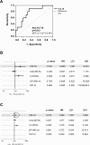
Fig 4. Effluent CCL18 concentrations predict the survival of the peritoneal membrane independent of classical transport parameters.
(A) Receiver operating characteristic (ROC) curve analysis of CCL18 effluent concentrations in samples from patients at 3 years of PD treatment who did or did not develop PMF. (AUC: area under the curve; CI: confidence interval). (B) Forest plot showing the hazard ratio (HR) and 95% upper and lower confidence intervals (UCI and LCI) of CCL18 and peritoneal membrane transport parameters in 35 patients measured at the third year of PD treatment. CCL18 showed significant association with PMF independent of U-MTAC, Cr -MTAC, or D/P Cr. (C) Forest plot showing the HR and 95% UCI and LCI of CCL18 and peritoneal membrane transport parameters measured at the last effluent determination in 61 patients who were treated for more than 3 years with PD. CCL18 showed a significant association with PMF, independent of U-MTAC, Cr-MTAC, or D/P Cr.
Fig 5
Fig 5. Time course analysis of PAI-1 values in patients treated with PD.
(A) Scatter plots showing mean, and SD values (B) Effluent PAI-1 values in patients who developed PMF (black bars; N = 10) or who did not (white bars; N = 33) Mean ±SD are shown. No significant differences were detected at any time point (Mann-Witney U test)
Fig 6
Fig 6. Cross-sectional study of PAI-1 values in 61 patients treated with PD with late (>3 years) peritoneal samples.
Scatter plots showing mean, and SD values (A) Significantly higher levels were found in effluent samples from the patients who developed PMF (p = .0038; Mann-Witney U test). (B) PAI-1 effluent concentrations in patients who developed EPS. No significant differences were detected (Mann-Witney U test)

References

    1. De Sousa-Amorim E, Del Peso G, Bajo MA, Alvarez L, Ossorio M, Gil F, et al. Can EPS development be avoided with early interventions? The potential role of tamoxifen—a single-center study. Perit Dial Int. 2014;34:582–593. doi: 10.3747/pdi.2012.00286 - DOI - PMC - PubMed
    1. Johnson DW, Cho Y, Livingston BE, Hawley CM, McDonald SP, Brown FG, et al. Encapsulating peritoneal sclerosis: incidence, predictors, and outcomes. Kidney Int. 2010;77:904–912. doi: 10.1038/ki.2010.16 - DOI - PubMed
    1. Del Peso G, Jiménez-Heffernan JA, Bajo MA, Aroeira LS, Aguilera A, Fernández-Perpén A, et al. Epithelial-to-mesenchymal transition of mesothelial cells is an early event during peritoneal dialysis and is associated with high peritoneal transport. Kidney Int. 2008;73 (Suppl 108):S26–S33. - PubMed
    1. Goodlad C, Tam FW, Ahmad S, Bhangal G, North BV, Brown EA. Dyalisate cytokine levels do not predict encapsuating peritoneal sclerosis. Perit Dial Int. 2014;34:594–604. doi: 10.3747/pdi.2012.00305 - DOI - PMC - PubMed
    1. Balasubramaniam G, Brown EA, Davenport A, Cairns H, Cooper B, Fan SL, et al. The Pan-Thames EPS study: treatment and outcomes of encapsulating peritoneal sclerosis. Nephrol Dial Transplant. 2009;24:3204–3215. - PubMed

LinkOut - more resources