. 2017 Apr 17;13(4):e1006338.
doi: 10.1371/journal.ppat.1006338. eCollection 2017 Apr.

The Epstein-Barr virus miR-BHRF1-1 targets RNF4 during productive infection to promote the accumulation of SUMO conjugates and the release of infectious virus

Affiliations

The Epstein-Barr virus miR-BHRF1-1 targets RNF4 during productive infection to promote the accumulation of SUMO conjugates and the release of infectious virus

Jinlin Li et al. PLoS Pathog. .

Abstract

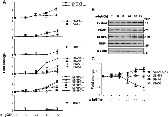
Post-translational modification by the Small Ubiquitin-like Modifier (SUMO) regulates a variety of cellular functions, and is hijacked by viruses to remodel the host cell during latent and productive infection. Here we have monitored the activity of the SUMO conjugation machinery in cells productively infected with Epstein-Barr virus (EBV). We found that SUMO2/3 conjugates accumulate during the late phase of the productive virus cycle, and identified several viral proteins as bone fide SUMOylation substrates. Analysis of the mechanism involved in the accumulation of SUMOylated proteins revealed upregulation of several components of the SUMO-conjugation machinery and post-transcriptional downregulation of the SUMO-targeted ubiquitin ligase RNF4. The latter effect was mediated by selective inhibition of RNF4 protein expression by the viral miR-BHRF1-1. Reconstitution of RNF4 in cells expressing an inducible miR-BHRF1-1 sponge or a miR-BHRF1-1 resistant RNF4 was associated with reduced levels of early and late viral proteins and impaired virus release. These findings illustrate a novel strategy for viral interference with the SUMO pathway, and identify the EBV miR-BHRF1-1 and the cellular RNF4 as regulators of the productive virus cycle.


Conflict of interest statement

The authors have declared that no competing interests exist.

Figures

Fig 1
Fig 1. SUMO conjugates accumulate during productive EBV infection.
Akata-Bx1 cells were harvested at the indicated times after induction by anti-IgG cross-linking and western blots were probed with antibodies to SUMO1 or SUMO2/3. (A) Induction of the productive cycle was confirmed in western blots probes with antibodies to the EBV transactivator BZLF1. Representative western blots illustrating the progressive accumulation of high molecular weight polypeptides reacting with antibodies to SUMO1 (B) and SUMO2/3 (C) antibodies; β-actin was used as loading control. * a longer exposure was required to visualize the free SUMO1.
Fig 2
Fig 2. The accumulation of SUMO conjugates is dependent on virus reactivation.
(A) Western blots of untreated and anti-IgG treated EBV negative Akata and EBV positive Akata-Bx1 and TPA/Bu treated EBV negative AGS and EBV positive AGS-Bx1 were probed with antibodies to SUMO2/3, BZLF1 and β-actin. Induction of the productive virus cycle was accompanied by accumulation of poly-SUMOylated proteins in the EBV positive cell lines. (B) The intensities of the SUMO2/3 reactive bands were quantified by densitometry. The average increase relative to untreated controls in two independent experiments is shown in the figure.
Fig 3
Fig 3. Expression of components of the SUMOylation machinery.
Protein and mRNA expression was monitored over time in induced Akata-Bx1 by qPCR and western blot. (A) Levels of specific mRNAs measured by qPCR. The results are shown as fold change relative to the expression at time 0. The mean ± SD of three or more experiments each performed in triplicate is shown. (B) Representative western blots illustrating the increased expression of SUMO2, PIAS1 and SENP6 and decreased expression of RNF4. β-actin was used as loading control. One representative western blot out of three where the proteins were tested in parallel is shown. (C) Quantification of protein expression. The mean ± SD of three experiments is shown. Statistical significance calculated by student’s T-test is indicated as: ** = p<0.001, *** = p<0.0001.
Fig 4
Fig 4. The downregulation of RNF4 is not due to increased protein turnover.
The western blot shown in Fig 2 was stripped and reprobed with RNF4 specific antibodies. (A) RNF4 is downregulated during productive infection in the EBV positive cell lines. (B) The intensity of the RNF4 specific band was quantified by densitometry and fold change of was calculated relative to untreated cells. The mean ± SD of two experiments is shown. (C) The productive virus cycle was induced in Akata-Bx1 cells by treatment with anti-IgG for 48 hrs, and one aliquot of the cells was treated with 10 μM of the proteasome inhibitor MG132 for 6 hrs before harvesting. A significant increase of the RNF4 specific band was observed in control cells following treatment with MG132, confirming that RNF4 is a bona fide proteasome substrate. Inhibition of the proteasome did not reverse the downregulation of RNF4 in the induced cells. (D) The intensity of the RNF4 specific band was quantified by densitometry and fold change was calculated relative to control DMSO treated cells. The mean ± SD of three experiments is shown.
Fig 5
Fig 5. The BHRF1-1 miRNA targets RNF4.
(A) HEK293T cells were co-transfected with an RNF4-3´UTR-LUC reporter plasmid and with plasmids expressing the indicated mature EBV miRNAs. The % luciferase activity relative to the control scrambled miRNA measured after 48h was: miR-BHRF1-1 0.65±0.03, mirBHRF1-2 1.01±0.08, miRBHRF1-3 0.92±0.06. The % decrease of luciferase activity was calculated relative to the luciferase activity of cells transfected with a control scrambled miRNA. Expression of miR-BHRF1-1 was reproducibly associated with a significant reduction of luciferase activity. The mean ± SD of three experiments is shown. (B) HEK293T cells were co-transfected with a plasmid expressing miR-BHRF1-1 and LUC-reporter plasmids containing wild type or mutant RNF4-3´UTR. Mutation of the miR-BHRF1-1 seed site was associated with loss of inhibition. The mean ± SD of three experiments is shown. (C) Representative western blot illustrating the expression of RNF4 in Akata-Bx1 cells transfected for 48 hrs with the indicated amount of a synthetic miR-BHRF1-1 oligonucleotide. (D) The expression of miR-BHRF1-1 in lentivirus transduced Akata-Bx1 was quantified by qPCR. A stable Akata-Bx1 subline expressing the inducible miRNA was culture in the presence or absence of doxycycline for 48 h. Mean ± SD of fold increase in three independent experiments each performed in triplicate. (E) Representative western blot illustrating the decrease of RNF4 in cells expressing miR-BHRF1-1. Cells transfected with a scrambled control miRNA were used as control and β-actin was used as loading control. One representative experiment out of three is shown. Statistical significance calculated by student’s T-test is indicated as: * = p<0.05, *** = p<0.0001.
Fig 6
Fig 6. Expression of a miR-BHRF1-1 sponge prevents the downregulation of RNF4.
The productive virus cycle was induced in Akata-Bx1 transduced with a recombinant lentivirus expressing a doxycycline regulated BHRF1-1 sponge. (A) Expression of the sponge and miR-BHRF1-1 was quantified by qPCR after 48 hrs. The upregulation of miR-BHRF1-1 was significantly reduced in cells expressing the sponge. The mean ± SD of three independent experiments is shown. (B) Total cell lysates were probed with a RNF4 specific antibody. The reduction of miR-BHRF1-1 in cells expressing the sponge prevented the downregulation of RNF4. One representative western blot out of three is shown. (C) Quantification of the RNF4 specific band in three independent experiments. Fold change was calculated relative to the expression of RNF4 in untreated Akata-Bx1.
Fig 7
Fig 7. The reconstitution of RNF4 expression prevents the accumulation of poly-SUMOylated proteins during productive infection.
The virus-reactivation induced downregulation of RNF4 was reversed by expression of a miR-BHRF1-1 sponge or by expression of a miR-BHRF1-1 resistant RNF4. (A) Expression of the sponge was associated with decreased of the SUMO conjugates. Western blots of total cell lysates harvested 48 hrs after induction were sequentially probed with antibodies specific for SUMO2/3 and β-actin. (B) Expression of a miR-BHRF1-1 resistant RNF4 prevents the accumulation of SUMO conjugates. Western blots of total cell lysates harvested 48 hrs after induction were probed with antibodies specific for RNF4, the FLAG tag, SUMO2/3 and β-actin. The transduced RNF4 is detected as a double band by the RNF4 specific antibody due to alternative translation that excludes the N-terminal tag. (C) Quantification of the western blots. The mean ± SD of two or more experiments is shown. Statistical significance calculated by student’s T-test is indicated as: * = p<0.05.
Fig 8
Fig 8. The decrease of SUMO conjugates in RNF4 reconstituted cells is due to proteasomal degradation.
Untreated and α-IgG treated Akata-Bx1 were cultured for 48 hrs and treated with the proteasome inhibitor MG132 for 6 hrs before harvesting. The decrease of SUMO conjugates induced by expression of the miR-BHRF1-1 sponge was reversed by inhibition of the proteasome. (A) One representative western blot out of three is shown. (B) The intensity of the SUMO2 reactive bands was quantified by densitometry. Fold change was calculated relative to untreated Akata-Bx1. Mean ± SD of three experiments. Statistical significance calculated by student’s T-test is indicated as: * = p<0.05.
Fig 9
Fig 9. Viral proteins are bona fide SUNO substrates.
Plasmids encoding FLAG-tagged version of the indicated viral proteins were co-transfected in HEK293T cells together with a plasmid expressing His-tagged SUMO2. Total cells lysates were probed with an anti-FLAG antibody (left panel). Cell pellets were lysed in denaturing buffer containing 7M urea and SUMOylated proteins were affinity purified with Ni-NTA agarose beads. Western blots were probed with the anti-FLAG antibody (right panel). High molecular weight species corresponding to poly-SUMOylated proteins were detected by the FLAG antibody. One representative experiment out of three is shown.
Fig 10
Fig 10. Reconstitution of RNF4 expression in productively infected cells is associated with proteasome-dependent degradation of viral proteins.
(A) Akata-Bx1 constitutively expressing the inducible miR-BHRF1-1 sponge were treated overnight with doxycycline and then induced by cross-linking surface IgG for 48 hrs. One aliquot of the cells was treated with the proteasome inhibitor MG132 during the last 6 hrs before harvesting. Western blots of total cell lysates were probed with the indicated antibodies. The downregulation of BGLF5 and BVRF2 observed in doxycycline treated cells was reversed by treatment with MG132, indicating that the proteins are degraded by the proteasome. One representative experiment out of two is shown. (B) The intensities of the specific bands were quantified by densitometry and fold change was calculated relative to cells induced in the absence of doxycycline. The mean ± SD of two experiments is shown.
Fig 11
Fig 11. Reconstitution of RNF4 hampers the release of virus particles.
The productive virus cycle was induced by anti-IgG treatment in Akata-Bx1, Akata-Bx1 expressing the inducible miR-BHRF1-1 sponge and Akata-Bx1 expressing a miR-BHRF1-1 resistant RNF4 cultured in the presence or absence of doxycycline. Intact virus particles released in the supernatants harvested after 3 and 7 days were measured by qPCR after treatment with DNase I. The amount of virus was estimated relative to a standard curve constructed by serial dilutions of a BZLF1 encoding plasmid. Reconstitution of RNF4 expression in cells expressing the miR-BHRF1-1 sponge (A) or miR-BHRF1-1 resistant RNF4 (B) was accompanied by a significantly reduced virus yield. The mean ± SD of two to three experiments is shown.

Similar articles

Cited by

References

    1. Flotho A, Melchior F. Sumoylation: a regulatory protein modification in health and disease. Annu Rev Biochem. 2013;82:357–85. 10.1146/annurev-biochem-061909-093311 - DOI - PubMed
    1. Manza LL, Codreanu SG, Stamer SL, Smith DL, Wells KS, Roberts RL, et al. Global shifts in protein sumoylation in response to electrophile and oxidative stress. Chem Res Toxicol. 2004;17(12):1706–15. 10.1021/tx049767l - DOI - PubMed
    1. Bossis G, Melchior F. Regulation of SUMOylation by reversible oxidation of SUMO conjugating enzymes. Mol Cell. 2006;21(3):349–57. 10.1016/j.molcel.2005.12.019 - DOI - PubMed
    1. Golebiowski F, Matic I, Tatham MH, Cole C, Yin Y, Nakamura A, et al. System-wide changes to SUMO modifications in response to heat shock. Sci Signal. 2009;2(72):ra24 10.1126/scisignal.2000282 - DOI - PubMed
    1. Desterro JM, Rodriguez MS, Kemp GD, Hay RT. Identification of the enzyme required for activation of the small ubiquitin-like protein SUMO-1. J Biol Chem. 1999;274(15):10618–24. - PubMed

MeSH terms

LinkOut - more resources