

Comparative expression profiling of testis-enriched genes regulated during the development of spermatogonial cells
- PMID: 28414809
- PMCID: PMC5393594
- DOI: 10.1371/journal.pone.0175787
Comparative expression profiling of testis-enriched genes regulated during the development of spermatogonial cells
Abstract
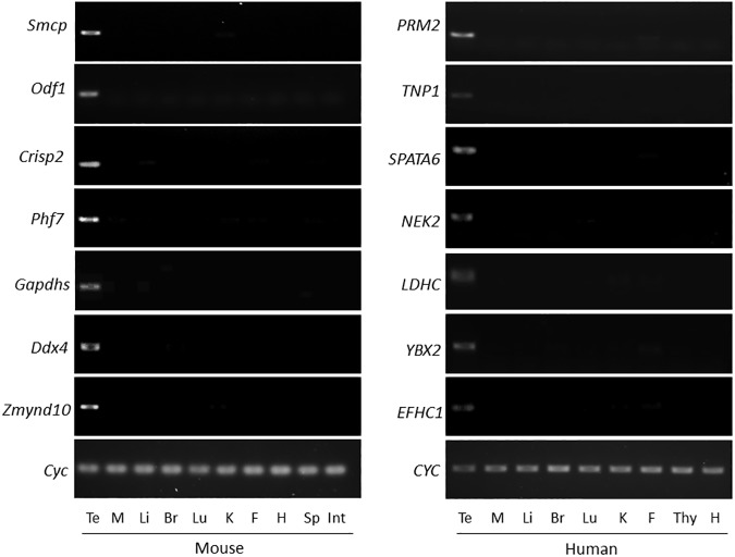
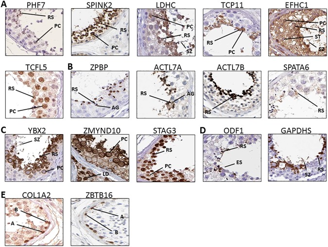
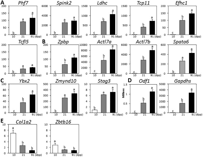
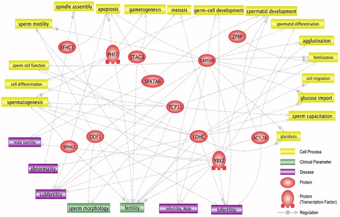
The testis has been identified as the organ in which a large number of tissue-enriched genes are present. However, a large portion of transcripts related to each stage or cell type in the testis still remains unknown. In this study, databases combined with confirmatory measurements were used to investigate testis-enriched genes, localization in the testis, developmental regulation, gene expression profiles of testicular disease, and signaling pathways. Our comparative analysis of GEO DataSets showed that 24 genes are predominantly expressed in testis. Cellular locations of 15 testis-enriched proteins in human testis have been identified and most of them were located in spermatocytes and round spermatids. Real-time PCR revealed that expressions of these 15 genes are significantly increased during testis development. Also, an analysis of GEO DataSets indicated that expressions of these 15 genes were significantly decreased in teratozoospermic patients and polyubiquitin knockout mice, suggesting their involvement in normal testis development. Pathway analysis revealed that most of those 15 genes are implicated in various sperm-related cell processes and disease conditions. This approach provides effective strategies for discovering novel testis-enriched genes and their expression patterns, paving the way for future characterization of their functions regarding infertility and providing new biomarkers for specific stages of spematogenesis.
Conflict of interest statement
Figures
Similar articles
-
Transcriptional regulation of P63 on the apoptosis of male germ cells and three stages of spermatogenesis in mice.Cell Death Dis. 2018 Jan 23;9(2):76. doi: 10.1038/s41419-017-0046-z. Cell Death Dis. 2018. PMID: 29362488 Free PMC article.
-
Gene expression study in the juvenile mouse testis: identification of stage-specific molecular pathways during spermatogenesis.Mamm Genome. 2006 Sep;17(9):956-75. doi: 10.1007/s00335-006-0029-3. Epub 2006 Sep 8. Mamm Genome. 2006. PMID: 16964443
-
Expression profiling of purified male germ cells: stage-specific expression patterns related to meiosis and postmeiotic development.Physiol Genomics. 2006 Jan 12;24(2):75-85. doi: 10.1152/physiolgenomics.00215.2004. Epub 2005 Nov 15. Physiol Genomics. 2006. PMID: 16291737
-
Spermatogenesis in mammals: proteomic insights.Syst Biol Reprod Med. 2012 Aug;58(4):179-90. doi: 10.3109/19396368.2012.691943. Syst Biol Reprod Med. 2012. PMID: 22788530 Review.
-
Roles of small ubiquitin-related modifiers in male reproductive function.Int Rev Cell Mol Biol. 2011;288:227-59. doi: 10.1016/B978-0-12-386041-5.00006-6. Int Rev Cell Mol Biol. 2011. PMID: 21482414 Review.
Cited by
-
Control of human testis-specific gene expression.PLoS One. 2019 Sep 12;14(9):e0215184. doi: 10.1371/journal.pone.0215184. eCollection 2019. PLoS One. 2019. PMID: 31514204 Free PMC article.
-
Actl7b deficiency leads to mislocalization of LC8 type dynein light chains and disruption of murine spermatogenesis.Development. 2023 Nov 1;150(21):dev201593. doi: 10.1242/dev.201593. Epub 2023 Oct 27. Development. 2023. PMID: 37800308 Free PMC article.
-
Pseudoautosomal Region 1 Overdosage Affects the Global Transcriptome in iPSCs From Patients With Klinefelter Syndrome and High-Grade X Chromosome Aneuploidies.Front Cell Dev Biol. 2022 Feb 3;9:801597. doi: 10.3389/fcell.2021.801597. eCollection 2021. Front Cell Dev Biol. 2022. PMID: 35186953 Free PMC article.
-
Adaptive Evolution and Functional Differentiation of Testis-Expressed Genes in Theria.Animals (Basel). 2024 Aug 9;14(16):2316. doi: 10.3390/ani14162316. Animals (Basel). 2024. PMID: 39199849 Free PMC article.
-
A novel testis-enriched gene, Samd4a, regulates spermatogenesis as a spermatid-specific factor.Front Cell Dev Biol. 2022 Oct 5;10:978343. doi: 10.3389/fcell.2022.978343. eCollection 2022. Front Cell Dev Biol. 2022. PMID: 36274854 Free PMC article.
References
-
- Uhlén M, Fagerberg L, Hallström BM, Lindskog C, Oksvold P, Mardinoglu A, et al. Proteomics. Tissue-based map of the human proteome. Science. 2015; 347: 1260419 doi: 10.1126/science.1260419 - DOI - PubMed
-
- Yu X, Lin J, Zack DJ, Qian J. Computational analysis of tissue-specific combinatorial gene regulation: predicting interaction between transcription factors in human tissues. Nucleic Acids Res. 2006; 34: 4925–4936. doi: 10.1093/nar/gkl595 - DOI - PMC - PubMed
-
- Ben-Tabou de-Leon S, Davidson EH. Gene regulation: gene control network in development. Annu Rev Biophys Biomol Struct. 2007; 36: 191–212. doi: 10.1146/annurev.biophys.35.040405.102002 - DOI - PubMed
-
- Djureinovic D, Fagerberg L, Hallström B, Danielsson A, Lindskog C, Uhlén M, et al. The human testis-specific proteome defined by transcriptomics and antibody-based profiling. Mol Hum Reprod. 2014; 20: 476–488. doi: 10.1093/molehr/gau018 - DOI - PubMed
-
- Griswold MD. The central role of Sertoli cells in spermatogenesis. Semin Cell Dev Biol. 1998; 9: 411–416. doi: 10.1006/scdb.1998.0203 - DOI - PubMed
Publication types
MeSH terms
Substances
Grants and funding
LinkOut - more resources
Full Text Sources
Other Literature Sources
Molecular Biology Databases
