. 2017 Apr 17;12(4):e0176000.
doi: 10.1371/journal.pone.0176000. eCollection 2017.

Isolation of human explant derived cardiac stem cells from cryopreserved heart tissue

Affiliations

Isolation of human explant derived cardiac stem cells from cryopreserved heart tissue

Robyn Jackson et al. PLoS One. .

Abstract

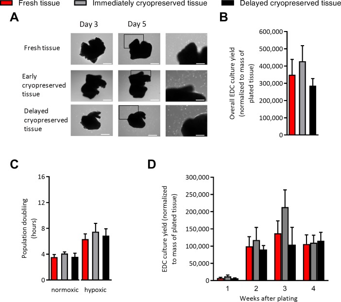
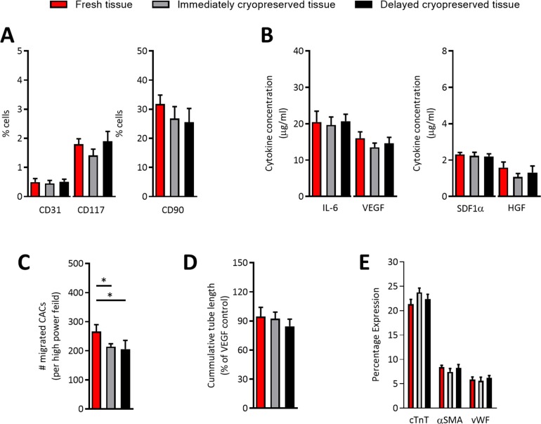
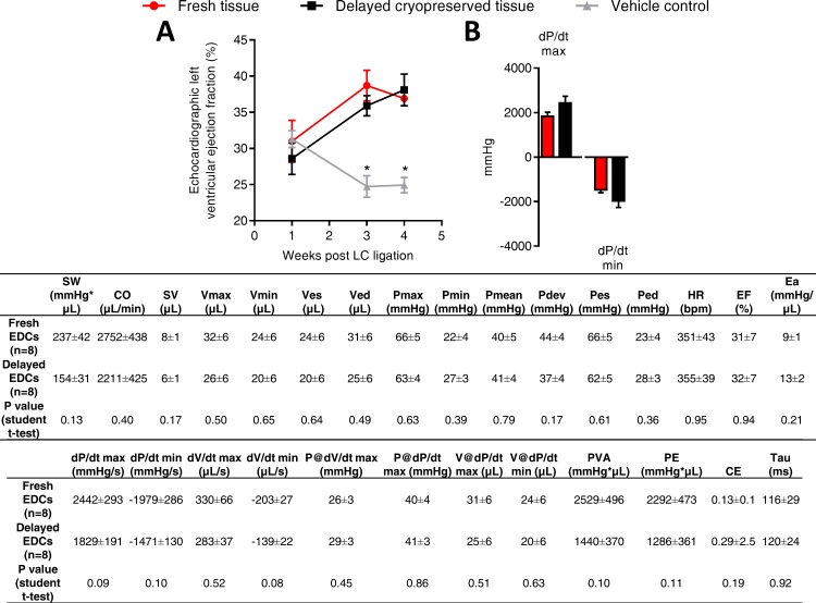
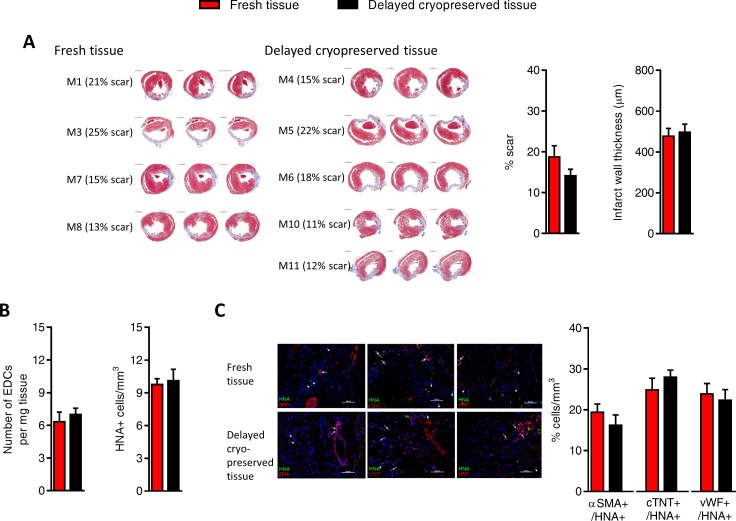
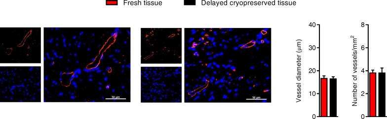
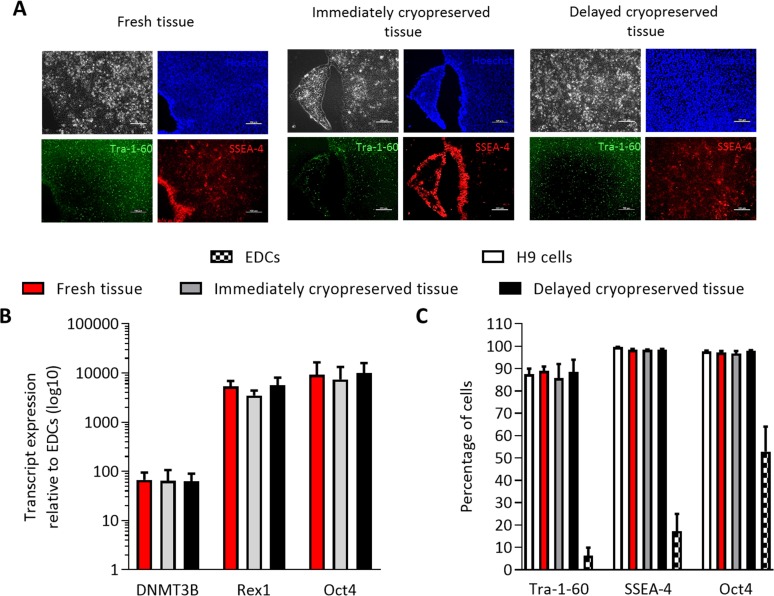
The value of preserving high quality bio specimens for fundamental research is significant as linking cellular and molecular changes to clinical and epidemiological data has fueled many recent advances in medicine. Unfortunately, storage of traditional biospecimens is limited to fixed samples or isolated genetic material. Here, we report the effect of cryopreservation of routine myocardial biopsies on explant derived cardiac stem cell (EDC) culture outcomes. We demonstrate that immediate cryopreservation or delayed cryopreservation after suspension within cardioplegia for 12 hours did not alter EDC yields, proliferative capacity, antigenic phenotype or paracrine signature. Cryopreservation had negligible effects on the ability of EDCs to adopt a cardiac lineage, stimulate new vessel growth, attract circulating angiogenic cells and repair injured myocardium. Finally, cryopreservation did not influence the ability of EDCs to undergo genetic reprogramming into inducible pluripotent stem cells. This study establishes a means of storing cardiac samples as a retrievable live cell source for cardiac repair or disease modeling.


Conflict of interest statement

Competing Interests: The authors have declared that no competing interests exist.

Figures

Fig 1
Fig 1. Effect of tissue cryopreservation on cell culture outcomes.
(A) Representative images of plated tissue with progressive growth of EDC from days 3 to 5. Left panels provide magnified portions of the middle panels to demonstrate cells spontaneously emerging to cover the cultureware. Left and middle panels size bar is 500 μm. Right panel size bar is 250 μm. (B) Effects of tissue cryopreservation on the overall numbers of EDCs cultured from atrial appendages normalized to the mass of the plated tissue (mean ± SEM, n = 5 explant cultures). (C) Effect of tissue cryopreservation on the proliferative capacity of EDCs plated in normoxic (21% oxygen; 20% serum) and hypoxic stress (1% oxygen; 1% serum) culture conditions (mean ± SEM, n = 5 explant cultures). (D) Cell culture yields taken at each weekly serial harvest from plated ventricular tissue using mild trypsinization normalized to mass of the plated tissue (mean ± SEM, n = 4 explant cultures).
Fig 2
Fig 2. Effects of cryopreservation on the in vitro profile of EDCs.
(A) Effects of tissue cryopreservation on the phenotype EDCs cultured from atrial appendages (mean ± SEM, n = 5 explant cultures). (B) Effects of tissue cryopreservation on the cytokine content within EDC conditioned media exposed to hypoxic (1% oxygen) low serum (1% serum) culture conditions (mean ± SEM, IL-6 = interleukin-6, HGF = hepatic growth factor, SDF1α = stromal cell derived factor 1α, VEGF = vascular endothelial growth factor, n = 5 conditioned media samples with 3 technical replicates). (C) Effects of tissue cryopreservation on the ability of EDC conditioned media to recruit circulating angiogenic cells (CACs) through transwell membrane (mean ± SEM, *p≤0.05, n = 5 conditioned media samples with 6 random fields/well). (D) Effects of tissue cryopreservation on the ability of EDC conditioned media to stimulate human umbilical vein endothelial cells tubule formation (mean ± SEM, n = 5 conditioned media samples). (D) Effect of tissue cryopreservation on the ability of EDCs to adopt a cardiomyocyte (cardiac troponin T, cTnT), endothelial (von Willebrand Factor, vWF) or smooth muscle (alpha smooth muscle actin, αSMA) lineage (mean ± SEM, n = 5 conditioned media samples).
Fig 3
Fig 3. Effect of cryopreservation on cell-mediated repair of ischemic myocardium.
(A) Effect of transplanting cryopreserved tissue sourced EDCs on echocardiographic function (mean ± SEM, *p≤0.05 vs. fresh or cryopreserved EDCs, n = 10–11 animals per group). (B) Effect of cryopreserved tissue sourced EDCs on invasive hemodynamics (mean ± SEM, n = 10–11 animals per group). SW, stroke work; CO, cardiac output; SV, stroke volume; Vmax, maximum volume; Vmin, minimum volume; Ves, end-systolic volume; Ved, end-diastolic volume; Pmax, maximum pressure; Pmin, minimum pressure; Pmean, mean pressure; Pdev, developed pressure; Pes, end-systolic pressure; Ped, end-diastolic pressure, HR, heart rate; EF, ejection fraction; Ea, arterial elastance; PowMax, maximum power; dP/dt max, maximum value of dP/dt; dP/dt min, minimum value of dP/dt; dV/dt max, maximum value of dV/dt; dV/dt min, minimum value of dV/dt min; P@dV/dt max, Pressure at maximum of dV/dt; P@dP/dt max, pressure at maximum of dP/dt max; V@dP/dt max, volume at maximum of dP/dt; V@dP/dt min, volume at minimum of dP/dt; PVA, pressure volume area; PE, potential energy; CE, cardiac efficiency; Tau, relaxation time constant calculated by Glantz method.
Fig 4
Fig 4. Effects of tissue cryopreservation on scar size, transplanted cell retention and transplanted cell fate.
(A) Representative images (left panel) and quantitative analysis (right panels) demonstrating that tissue cryopreservation had no discernable effect on the ability of transplanted EDCs to reduce left ventricular scar sizes (mean ± SEM, n = 4–5 animals per group with 3 histological slides analyzed per animal, scale bar 1000 μm). (B) Quantitative PCR for retained human alu sequences and histological analysis for human nuclear antigen positive (HNA+) cells demonstrating that cryopreservation had no effect on transplanted EDCs engraftment (mean ± SEM, n = 5–6 animals per group). (C) Representative images of immunohistochemical sections used for peri-infarct field quantification of cell engraftment and co-localization with markers of cardiac myocyte (cTNT, upper panel), smooth muscle (αSMA, middle panel) and endothelial (vWF, lower panel) lineage (scale bar 50 μm). Arrows indicate examples of HNA co-segregation with lineage markers while arrow heads indicate HNA staining alone. Histological analysis demonstrating the co-segregation between HNA+ cells and markers of transplanted cell lineage commitment (mean ± SEM, n = 6 animals per group).
Fig 5
Fig 5. Effects of tissue cryopreservation on post-infarct neo-vascularization.
Representative images (left panel) and quantitative analysis (right panels) demonstrating that tissue cryopreservation had no effect on the ability of transplanted EDCs to promote new vessel growth (mean ± SEM, n = 4–5 animals per group with 3 histological slides analyzed per animal, scale bar 50 μm).
Fig 6
Fig 6. Effect of tissue cryopreservation on induced pluripotent stem cell reprogramming of EDCs.
(A) Representative images of live iPSC colonies that express markers of pluripotent identity (SSEA-4 and Tra-1-60). Size bar is 100 μm. (B) Effect of tissue cryopreservation on pluripotent transcript expression of iPSC reprogrammed EDCs (mean ± SEM, n = 3 cell lines). (C) Effect of tissue cryopreservation on flow cytometry expression of pluripotent surface antigens (mean ± SEM, n = 3 cell lines).

References

    1. Baker M. Biorepositories: Building better biobanks. Nature. 2012;486(7401):141–6. doi: 10.1038/486141a - DOI - PubMed
    1. Eiseman E, Haga S. A handbook of human tissue sources: A national resource of human tissue samples. Santa Monica, CA: 2000.
    1. Yokomuro H, Mickle DA, Weisel RD, Li RK. Optimal conditions for heart cell cryopreservation for transplantation. Mol Cell Biochem. 2003;242(1–2):109–14. - PubMed
    1. Makkar RR, Smith RR, Cheng K, Malliaras K, Thomson LE, Berman D, et al. Intracoronary cardiosphere-derived cells for heart regeneration after myocardial infarction (CADUCEUS): a prospective, randomised phase 1 trial. Lancet. 2012;379(9819):895–904. doi: 10.1016/S0140-6736(12)60195-0 - DOI - PMC - PubMed
    1. Molgat AS, Tilokee EL, Rafatian G, Vulesevic B, Ruel M, Milne R, et al. Hyperglycemia inhibits cardiac stem cell-mediated cardiac repair and angiogenic capacity. Circulation. 2014;130(11 Suppl 1):S70–S6. - PubMed