. 2017 May 9;8(19):31626-31637.
doi: 10.18632/oncotarget.16382.

Inhibition of iron overload-induced apoptosis and necrosis of bone marrow mesenchymal stem cells by melatonin

Affiliations

Inhibition of iron overload-induced apoptosis and necrosis of bone marrow mesenchymal stem cells by melatonin

Fan Yang et al. Oncotarget. .

Abstract

Iron overload induces severe damage to several vital organs such as the liver, heart and bone, and thus contributes to the dysfunction of these organs. The aim of this study is to investigate whether iron overload causes the apoptosis and necrosis of bone marrow mesenchymal stem cells (BMSCs) and melatonin may prevent its toxicity. Perls' Prussion blue staining showed that exposure to increased concentrations of ferric ammonium citrate (FAC) induced a gradual increase of intracellular iron level in BMSCs. Trypan blue staining demonstrated that FAC decreased the viability of BMSCs in a concentration-dependent manner. Notably, melatonin protected BMSCs against apoptosis and necrosis induced by FAC and it was vertified by Live/Dead, TUNEL and PI/Hoechst stainings. Furthermore, melatonin pretreatment suppressed FAC-induced reactive oxygen species accumulation. Western blot showed that exposure to FAC resulted in the decrease of anti-apoptotic protein Bcl-2 and the increase of pro-apoptotic protein Bax and Cleaved Caspase-3, and necrosis-related proteins RIP1 and RIP3, which were significantly inhibited by melatonin treatment. At last, melatonin receptor blocker luzindole failed to block the protection of BMSCs apoptosis and necrosis by melatonin. Taken together, melatonin protected BMSCs from iron overload induced apoptosis and necrosis by regulating Bcl-2, Bax, Cleaved Caspase-3, RIP1 and RIP3 pathways.

Keywords: apoptosis; bone marrow mesenchymal stem cells; iron overload; melatonin; necrosis.


Conflict of interest statement

CONFLICTS OF INTEREST

The authors had no conflicts of interest to declare in relation to this article.

Figures

Figure 1
Figure 1. Iron overload caused the decrease of cell viability in BMSCs
(A) Perls’ Prussion blue staining was applied to detect intracellular iron accumulation in FAC-induced BMSCs. After exposure to FAC 50 μM, 100 μM, 200 μM and 400 μM, intracellular iron level was gradually increased. (B) Flow cytometry analysis showed that treatment with FAC resulted in a concentration-dependent decrease of live BMSCs. (C) Trypan blue exclusion assay showed that exposure of BMSCs to FAC gradually decreased the number of live cells and increased the number of dead cells. Values are the mean ± SEM of 3 independent experiments (n = 3). **p < 0.01 versus Ctrl; ***p < 0.001 versus Ctrl.
Figure 2
Figure 2. Protective effect of melatonin on FAC-induced cell death of BMSCs
Live/Dead assay was applied to observe the effects of melatonin on FAC-induced cell death of BMSCs. Exposure of BMSCs to FAC 200 μM induced an increase of dead cells (red) compared to control group. But pretreatment with melatonin 100 μM abolished FAC-induced the increase of dead BMSCs. Data are for three independent experiments (n = 3).
Figure 3
Figure 3. Melatonin protected BMSCs against FAC-induced apoptosis and necrosis
(A) TUNEL staining showed that exposure of BMSCs to FAC 200 μM markedly increased the number of apoptotic positive cells compared to the control group. However, melatonin 100 μM led to a significant decrease in the number of apoptotic cells in FAC-induced BMSCs. (B) PI/Hoechst staining showed that exposure of BMSCs to FAC 200 μM for 24 h induced an increase of Hoechst-positive and PI-positive BMSCs. Afterwards, pretreatment with melatonin 100 μM countered the increase of apoptotic and necrotic positive cells induced by FAC 200 μM. Data are for three independent experiments (n = 3). ###p < 0.001, FAC 200 μM versus Ctrl; *p < 0.05, **p < 0.01, ***p < 0.001, FAC 200 μM + melatonin 100 μM versus FAC 200 μM.
Figure 4
Figure 4. Melatonin protected BMSCs against FAC-induced intracellular ROS increase
H2DCF-DA probe was applied to assessed the intracellular ROS production in FAC-induced BMSCs. ROS staining showed that exposed to FAC 200 μM for 24 h, BMSCs exhibited a gradual increase of intracellular ROS generation as illuminated by enhanced green staining in the cytoplasm. However, melatonin 100 μM suppressed the increase of ROS-positive cells with FAC treatment. Data are for three independent experiments (n = 3).
Figure 5
Figure 5. Melatonin suppressed FAC-induced BMSCs death by regulating Bcl-2/Bax and RIP pathways
Western blot showed that the exposure of BMSCs to FAC 200 μM caused a significant downregulation of Bcl-2 and upregulation of Bax, Cleaved Caspase-3, RIP1 and RIP3 proteins. However, pretreatment with melatonin 100 μM suppressed the decreased expression of Bcl-2, and the increased expression of Bax, Cleaved Caspase-3, RIP1 and RIP3 in FAC-treated BMSCs. Values are the mean ± SEM of 3 independent experiments (n = 3). ##p < 0.01, ###p < 0.001, FAC 200 μM versus Ctrl, *p < 0.05, **p < 0.01, FAC 200 μM + melatonin 100 μM versus FAC 200 μM.
Figure 6
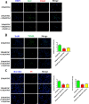
Figure 6. Effects of melatonin inhibitor luzindole on FAC-induced BMSCs death
(A) Live/Dead staining showed that melatonin 100 μM induced the decrease of dead cells with red staining, which can not be abrogated by the melatonin receptor blocker luzindole 10 μM. (B) TUNEL staining confirmed that FAC 200 μM led to significant increase in the number of TUNEL positive cells, which could be obviously reversed by melatonin 100 μM. However, luzindole failed to suppress the protective function of melatonin in FAC-treated BMSCs. (C) PI/Hoechst staining showed that luzindole 10 μM failed to block the protection of melatonin 100 μM on FAC-induced apoptosis and necrosis of BMSCs. Data are for three independent experiments (n = 3). *p < 0.05, ***p < 0.001, FAC 200 μM + melatonin 100 μM versus FAC 200 μM.
Figure 7
Figure 7. Luzindole has no effects on apoptotic and necrotic proteins in BMSCs
Western blot showed that the expression of Bax, Cleaved Caspase-3, RIP1 and RIP3 were significantly decreased by melatonin 100 μM. The increased expression of Bcl-2, decreased expression of Bax, Cleaved Caspase-3, RIP1 and RIP3 in melatonin pretreatment were not reversed by luzindole 10 μM. Values are the mean ± SEM of 3 independent experiments (n = 3). ***p < 0.001, FAC 200 μM + melatonin 100 μM versus FAC 200 μM.

Similar articles

Cited by

References

    1. Sekiya I, Larson BL, Vuoristo JT, Cui JG, Prockop DJ. Adipogenic differentiation of human adult stem cells from bone marrow stroma (MSCs) J Bone Miner Res. 2004;19:256–264. - PubMed
    1. Yang YJ, Qian HY, Huang J, Geng YJ, Gao RL, Dou KF, Yang GS, Li JJ, Shen R, He ZX, Lu MJ, Zhao SH. Atorvastatin treatment improves survival and effects of implanted mesenchymal stem cells in post-infarct swine hearts. Eur Heart J. 2008;29:1578–1590. - PubMed
    1. Wang A, Shen F, Liang Y, Wang J. Marrow-derived MSCs and atorvastatin improve cardiac function in rat model of AMI. Int J Cardiol. 2011;150:28–32. - PubMed
    1. Dong Q, Yang Y, Song L, Qian H, Xu Z. Atorvastatin prevents mesenchymal stem cells from hypoxia and serum-free injury through activating AMP-activated protein kinase. Int J Cardiol. 2011;153:311–316. - PubMed
    1. Noronha-Matos JB, Coimbra J, Sa-e-Sousa A, Rocha R, Marinhas J, Freitas R, Guerra-Gomes S, Ferreirinha F, Costa MA, Correia-de-Sa P. P2×7-induced zeiosis promotes osteogenic differentiation and mineralization of postmenopausal bone marrow-derived mesenchymal stem cells. FASEB J. 2014;28:5208–5222. - PubMed

MeSH terms