The N-terminal polypeptide derived from viral macrophage inflammatory protein II reverses breast cancer epithelial-to-mesenchymal transition via a PDGFRα-dependent mechanism
- PMID: 28415580
- PMCID: PMC5514921
- DOI: 10.18632/oncotarget.16394
The N-terminal polypeptide derived from viral macrophage inflammatory protein II reverses breast cancer epithelial-to-mesenchymal transition via a PDGFRα-dependent mechanism
Abstract
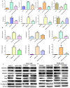
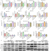
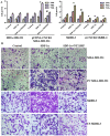
NT21MP, a 21-residue peptide derived from the viral macrophage inflammatory protein II, competed effectively with the natural ligand of CXC chemokine receptor 4 (CXCR4), stromal cell-derived factor 1-alpha, to induce apoptosis and inhibit growth in breast cancer. Its role in tumor epithelial-to-mesenchymal transition (EMT) regulation remains unknown. In this study, we evaluated the reversal of EMT upon NT21MP treatment and examined its role in the inhibition of EMT in breast cancer. The parental cells of breast cancer (SKBR-3 and MCF-7) and paclitaxel-resistant (SKBR-3 PR and MCF-7 PR) cells were studied in vitro and in combined immunodeficient mice. The mice injected with SKBR-3 PR cells were treated with NT21MP through the tail vein or intraperitoneally with paclitaxel or saline. Sections from tumors were evaluated for tumor weight and EMT markers based on Western blot. In vitro, the effects of NT21MP, CXCR4 and PDGFRα on tumor EMT were assessed by relative quantitative real-time reverse transcription-polymerase chain reaction, western blot and biological activity in breast cancer cell lines expressing high or low levels of CXCR4. Our results illustrated that NT21MP could reverse the phenotype of EMT in paclitaxel-resistant cells. Furthermore, we found that NT21MP governed PR-mediated EMT partly due to controlling platelet-derived growth factors A and B (PDGFA and PDGFB) and their receptor (PDGFRα). More importantly, NT21MP down-regulated AKT and ERK1/2 activity, which were activated by PDGFRα, and eventually reversed the EMT. Together, these results indicated that CXCR4 overexpression drives acquired paclitaxel resistance, partly by activating the PDGFA and PDGFB/PDGFRα autocrine signaling loops that activate AKT and ERK1/2. Inhibition of the oncogenic EMT process by targeting CXCR4/PDGFRα-mediated pathways using NT21MP may provide a novel therapeutic approach towards breast cancer.
Keywords: PDGFRα; breast cancer; epithelial-mesenchymal transition; paclitaxel; viral macrophage inflammatory protein II.
Conflict of interest statement
The authors declare that they have no conflicts of interest.
Figures


Similar articles
-
NT21MP negatively regulates paclitaxel-resistant cells by targeting miR‑155‑3p and miR‑155-5p via the CXCR4 pathway in breast cancer.Int J Oncol. 2018 Sep;53(3):1043-1054. doi: 10.3892/ijo.2018.4477. Epub 2018 Jul 9. Int J Oncol. 2018. PMID: 30015868 Free PMC article.
-
Antitumour activity of the recombination polypeptide GST-NT21MP is mediated by inhibition of CXCR4 pathway in breast cancer.Br J Cancer. 2014 Mar 4;110(5):1288-97. doi: 10.1038/bjc.2014.1. Epub 2014 Jan 21. Br J Cancer. 2014. PMID: 24448360 Free PMC article.
-
N-peptide of vMIP-Ⅱ reverses paclitaxel-resistance by regulating miRNA-335 in breast cancer.Int J Oncol. 2017 Sep;51(3):918-930. doi: 10.3892/ijo.2017.4076. Epub 2017 Jul 19. Int J Oncol. 2017. PMID: 28731125
-
Targeting the Molecules in EMT: A Potential Therapeutic Opportunity in Breast Cancer.Curr Mol Med. 2025;25(5):567-588. doi: 10.2174/0115665240310780240805114133. Curr Mol Med. 2025. PMID: 39162281 Review.
-
Anticancer effects of Traditional Chinese Medicine on epithelial-mesenchymal transition(EMT) in breast cancer: Cellular and molecular targets.Eur J Pharmacol. 2021 Sep 15;907:174275. doi: 10.1016/j.ejphar.2021.174275. Epub 2021 Jun 30. Eur J Pharmacol. 2021. PMID: 34214582 Review.
Cited by
-
The N-terminal polypeptide derived from vMIP-II exerts its anti-tumor activity in human breast cancer by regulating lncRNA SPRY4-IT1.Biosci Rep. 2018 Oct 17;38(5):BSR20180411. doi: 10.1042/BSR20180411. Print 2018 Oct 31. Biosci Rep. 2018. PMID: 30104400 Free PMC article.
-
Protocol for spatial analysis of multiple markers across adjacent tissue sections captured by RNAscope using CellProfiler.STAR Protoc. 2025 Jun 20;6(3):103914. doi: 10.1016/j.xpro.2025.103914. Online ahead of print. STAR Protoc. 2025. PMID: 40544447 Free PMC article.
-
Identified the novel resistant biomarkers for taxane-based therapy for triple-negative breast cancer.Int J Med Sci. 2021 Apr 26;18(12):2521-2531. doi: 10.7150/ijms.59177. eCollection 2021. Int J Med Sci. 2021. PMID: 34104083 Free PMC article.
-
NT21MP negatively regulates paclitaxel-resistant cells by targeting miR‑155‑3p and miR‑155-5p via the CXCR4 pathway in breast cancer.Int J Oncol. 2018 Sep;53(3):1043-1054. doi: 10.3892/ijo.2018.4477. Epub 2018 Jul 9. Int J Oncol. 2018. PMID: 30015868 Free PMC article.
-
The Signaling Duo CXCL12 and CXCR4: Chemokine Fuel for Breast Cancer Tumorigenesis.Cancers (Basel). 2020 Oct 21;12(10):3071. doi: 10.3390/cancers12103071. Cancers (Basel). 2020. PMID: 33096815 Free PMC article. Review.
References
-
- Fan L, Strasser-Weippl K, Li JJ, St Louis J, Finkelstein DM, Yu KD, Chen WQ, Shao ZM, Goss PE. Breast cancer in China. Lancet Oncol. 2014;15:279–289. - PubMed
-
- Siegal R, Naishadham D, Jemal A. Cancer Statistics. CA Cancer J Clin. 2013;2013(63):11–30. - PubMed
-
- NCCN Clinical . Practice Guidelines in Oncology (NCCN Guidelines), Breast Cancer. Version 3.2014 www.NCCN.org/patients.
-
- De Lena M, Latorre A, Calabrese P, Catino A, Lorusso V, Mazzei A, Aloe A. High efficacy of paclitaxel and doxorubicin as first-line therapy in advanced breast cancer: a phase I-II study. J Chemother. 2000;12:367–373. - PubMed
-
- Sabbah M, Emami S, Redeuilh G, Julien S, Prévost G, Zimber A, Ouelaa R, Bracke M, De Wever O, Gespach C. Molecular signature and therapeutic perspective of the epithelial-to-mesenchymal transitions in epithelial cancers. Drug Resist Updat. 2008;11:123–151. - PubMed
MeSH terms
Substances
LinkOut - more resources
Full Text Sources
Other Literature Sources
Medical
Research Materials
Miscellaneous

