. 2017 May 9;8(19):31041-31056.
doi: 10.18632/oncotarget.16055.

Inhibitor SBFI26 suppresses the malignant progression of castration-resistant PC3-M cells by competitively binding to oncogenic FABP5

Affiliations

Inhibitor SBFI26 suppresses the malignant progression of castration-resistant PC3-M cells by competitively binding to oncogenic FABP5

Waseem Al-Jameel et al. Oncotarget. .

Erratum in

Abstract

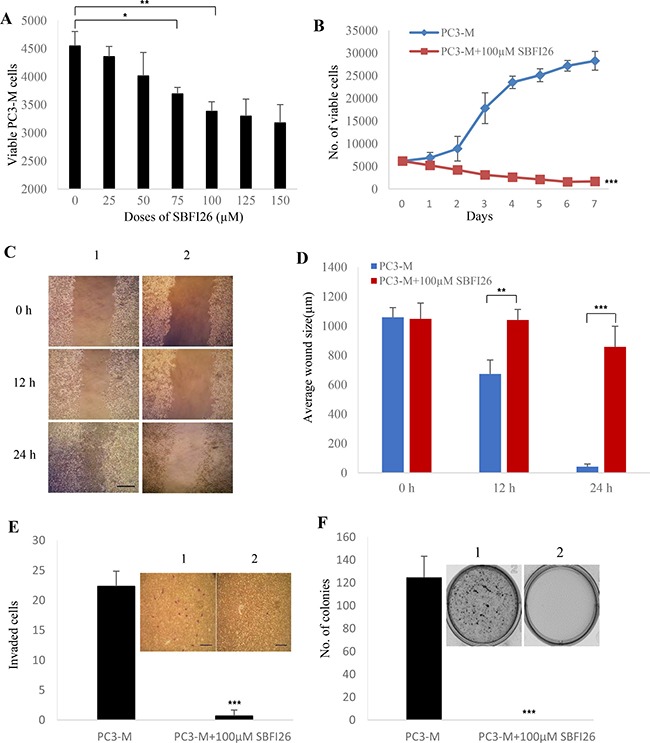
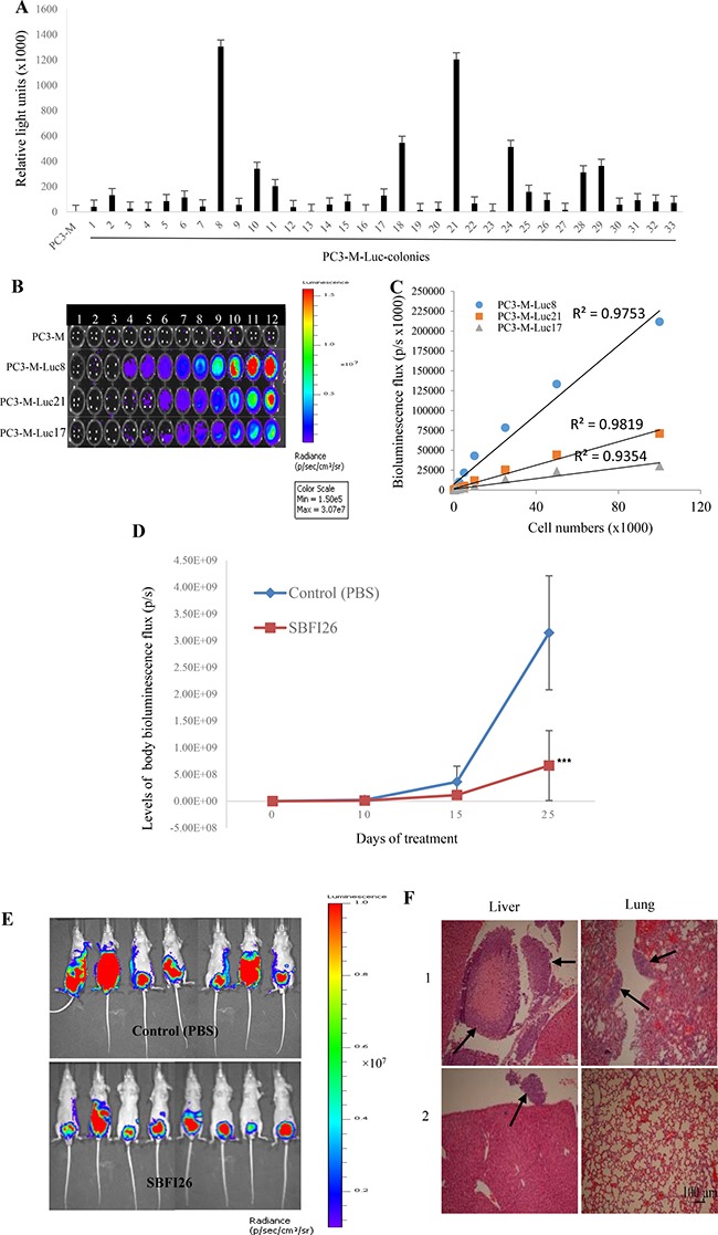
Castration resistant-prostate cancer is largely impervious to feather hormonal therapy and hence the outlook for patients is grim. Here we use an approach to attach the recently discovered Achilles heel. The experimental treatment established in this study is based on the recent discovery that it is the FABP5-PPARγ-VEGF signalling axis, rather than the androgen receptor pathway, played a dominant role in promoting the malignant progression of castration resistant prostate cancer cells. Treatments have been established in mice by suppressing the biological activity of FABP5 using a chemical inhibitor SBFI26. The inhibitor significantly suppressed the proliferation, migration, invasiveness and colony formation of PC3-M cells in vitro. It also produced a highly significant suppression of both the metastases and the primary tumours developed from cancer cells implanted orthotopically into the prostate glands of the mice. The inhibitor SBFI26 interferes with the FABP5-PPARγ- signalling pathway at the initial stage of the signal transduction by binding competitively to FABP5 to inhibit cellular fatty acid uptake. This avoids the fatty-acid stimulation of PPARγ and prevents it activating the down-stream regulated cancer-promoting genes. This entirely novel experimental approach to treating castration- resistant prostate cancer is completely different from current treatments that are based on androgen-blockade therapy.

Keywords: CRPC; FABP5; PPARγ; SBFI26; metastasis.


Conflict of interest statement

CONFLICTS OF INTEREST

The authors declare no potential conflicts of interest.

Figures

Figure 1
Figure 1. Detection of binding affinities of 4 candidate compounds to wtrFABP5 with DAUDA displacement assay to identify the lead chemical inhibitor of FABP5
(A) Chart records of binding affinity analysis of 3 different fatty acids and 4 different candidate chemical inhibitors of FABP5. a) Titration curve of DAUDA binding to wtrFABP5. Fixed amounts (3 μM) of wtrFABP5 were incubated with increasing concentrations of DAUDA (0.4–3 μM). For calculation of the dissociation constant Kd values, the excitation and emission used was 345 and 530 nm, respectively and the fluorescence data was normalized to the peak fluorescent intensity for each experiment and data of samples without protein was subtracted. The data was fitted by nonlinear regression techniques using GraphPad Prism software to a saturation binding curve model to estimate the apparent dissociation constant (Kd) and maximal fluorescence intensity (Bmax). The apparent dissociation constant (Kd) for DAUDA to wtrFABP5 was calculated to be 1.86 ± 0.16 μM. (b, c, d) Inhibition constant Ki (binding affinity) of Linoleic, Oleic and Palmitic acids binding to wtrFABP5. The Ki was measured to determine the potency of binding of these fatty acids with wtrFABP5 by evaluating their ability to displace DAUDA. The data were collected by displacement of 2 μM DAUDA from 3 μM wtrFABP5 in the presence of different concentrations of each fatty acid (0.5–20 μM). All data were fitted to a one site binding affinity model by non-linear regression techniques using GraphPad Prism software to estimate the binding affinity. The Ki of each ligand was determined using the equation Ki = IC50/1+ (DAUDA concentration /Kd). The binding affinity of Linoleic acid (Ki = 1.57 μM) was higher than that of Oleic and Palmitic acids (Ki = 1.88 and 4.30 μM, respectively). (e, f, g) Inhibition constant Ki of SBFI26, SBFI19, SBFI27, and SBFI31 to wtrFABP5. The data were collected by displacement of 2 μM DAUDA from 3μM wtrFABP5 in the presence of different concentrations (0.5–20 μM) of each chemical compound. All data were fitted to a one site binding affinity model by non-linear regression techniques using GraphPad Prism software to estimate the binding affinity. The binding affinity of SBFI26 (Ki =1.68 μM) was the highest amongst the 4 compounds. (B) Ki values of 3 different fatty acids and 4 different candidate chemical inhibitors. (C) Fluorescence intensity of displacement of 2 μM DAUDA binding from 3μM wtrFABP5 in the presence of 10 μM of 3 different fatty acids and 4 different candidate chemical inhibitors. The value of fluorescence intensity produced by the buffer and DAUDA plus wtrFABP5 was set as control. The results (mean ± SE) were obtained from 3 separate experiments (2-tailed unpaired Student’s t test, ***P < 0.0001).
Figure 2
Figure 2. Inhibitory effect of SBFI26 on proliferation, migration, invasion and anchorage-independent growth of the androgen-independent PC3-M prostate cancer cells
(A) Determination of the optimal inhibitory concentration of SBFI26 at which the maximum suppression of cell growth is achieved. MTT assay was performed to measure the viable PC3-M cell numbers of the control (untreated) and those treated with different concentrations of SBFI26 for 24 h. (B) Inhibitory effect of 100 μM SBFI26 on proliferation of PC3-M cells over the 7day experimental period. (C) Representative photos of the wound healing assay. PC3-M cells were grown in 6-well plates to form a monolayer. Scratches were made using 1 mL sterile pipette tip. Cell migration capacity was measured by the reduction in wound size in control (1) and in cultures treated with 100 μM SBFI26 (2) observed at 0, 12 and 24 hours after treatment. The scale bar is 250 μm. (D) Average wound sizes (μm) of the control PC3-M and cultures treated with 100 μM SBFI26 observed at 0, 12 and 24 hours after treatment. Data was collected by measuring image of the wound space and analyzed by ImageJ software (National Institutes of Health). (E) Number of invading cells from the control PC3-M cells (1) and cultures treated with 100 μM SBFI26 (2) for 24 h after different treatments. Results (mean ± SE) are obtained from three separate measurements. Scale bar is 250 μm. (F) Colonies produced by the control PC3-M cells (1) and cultures treated with 100 μM SBFI26 (2) in soft agar 2 weeks after the different treatments. Results (mean ± SE) are obtained from three separate plates in each treatment. The inserted picture was a representative plate from each of the 3 treatments. All in vitro results were subjected to 2-tailed unpaired Student’s t test and *P < 0.05; **P < 0.001; ***P < 0.0001.
Figure 3
Figure 3. Inhibitory effect of SBFI26 on tumorigenicity and metastatic ability of PC3-M cells implanted orthotopically into the prostate gland of the nude mouse
(A) Establishment of stable PC3-M colonies expressing strong bioluminescence signals by pGL4.50 [luc2/CMV/Hygro] vector transfection. Relative light units (mean ± SE) of the PC3-M parental cells and 33 colonies derived from PC3-M cells were obtained from 3 separate measurements. Individual colonies were isolated by ring cloning and 3 colonies that stably-expressed the highest bioluminescence signals were identified using D-luciferin (Promega) with a Varioskan Flash Reader (Thermo Scientific). (B) Detailed observation of the intensities of the bioluminescence images of the serially-diluted (20-100000) parental PC3-M cells and 3 representative PC3M-Luc transfectants. Association of the luminescence intensity with the number of cells was assessed by an IVIS imaging system (Perkin Elmer). The color bar on the right indicates the signal intensity range (photons/second/cm2). (C) Correlation between the bioluminescence flux intensity (photons/second) and the number of cells derived from 3 different PC3-M-Luc colonies. (D) Whole body tumor bioluminescence flux produced by each group of nude mice after orthotopic implantation of luciferase-labelled PC3-M cells and treated with PBS (control), SBFI26 (1 mg/kg) for 25 days. Values were plotted as mean ± SE (error bars) (n = 8); the difference between the control and each of the testing groups was assessed by two-tailed unpaired Student’s t test ***P < 0.0001. (E) Ventral bioluminescence images of primary tumors and metastases in all groups of experimental mice 25 days after treatment. The color bar on the right indicates the signal intensity range (photons/second/cm2). (F) Representative photomicrographs of detection of liver and lung metastases (arrows) from mice which received injection of PBS (1) and SBFI26 (2). Sections of tissues were stained with H&E. Magnification, ×10 and scale bar is 100 μm. All animal work was performed in accordance with UKCCCR guidelines under Home Office License PPL40/2963.
Figure 4
Figure 4. Effect of SBFI26 or GW9662 on tumorigenicity in prostate cancer xenograft mice
(A) Average volume of tumors produced by each group of male nude mice after subcutaneous inoculation of PC3-M cells (2 × 10⁶) and treated with PBS (control) or SBFI26 (1 mg/kg) for 31 days; started on day 1 and day 7 after inoculation. Values are plotted as mean ± SE (error bars) (n = 8); difference between the control group and the experimental groups were assessed by 2-tailed unpaired Student’s t test, **P < 0.001. (B) Representative mouse and its corresponding tumors from control (1) and SBFI26 treated (2) groups. (C) Average weight (mg) of tumours from control and SBFI26 treated groups of mice. Values were plotted as mean ± SE (error bars). The differences between the control and the experimental groups were assessed by 2-tailed unpaired Student’s t test **P < 0.001. (D) Average volume of tumors produced by each group of male nude mice after subcutaneous inoculation with PC3-M cancer cells and treated with PBS (control) and PPARγ antagonist (GW9662;1 mg/kg) for 31 days. Values were plotted as mean ± SE (error bars) (n = 5); differences between the control and the experimental groups were assessed by 2-tailed unpaired Student’s t test ***P < 0.0001. (E) Representative mouse and its corresponding tumor from each of the control (1) and GW9662 (2) groups. (F) Average weight (mg) of tumours in the control and experimental groups of mice. Values were plotted as mean ± SE (error bars). Differences between the control and the experimental groups were assessed by 2-tailed unpaired Student’s t test ***P < 0.0001.
Figure 5
Figure 5. Fatty acid uptake of different prostate epithelial cell lines and inhibitory effect of SBFI26 in PC3-M cells
(A) Representative histograms for unstained PNT2, LNCaP, 22RVI and PC3-M cells without adding BODIPY-labelled fatty acid. The marker M1 highlights negative peaks of the subclass control. (B) Representative histograms for fluorescence of stained PNT2, LNCaP, 22RVI and PC3-M cells 30min after adding BODIPY-labelled fatty acid and the marker M2 is placed to the right of M1 to highlight positive events (total percentage of cells with BODIPY-labeled fatty acid). (C) Percentages of cells taking up BODIPY-labelled fatty acid from different prostate epithelial cell lines. (D) Representative histograms for fatty acid uptake of PC3-M cells at a fixed concentration of BODIPY-labelled fatty acid with different concentrations of SBFI26. M1, unstained cells; M2, stained cells. (E) Percentages of cells with fatty acid uptake from PC3-M control (untreated) and those treated with different concentrations of SBFI26 for 30min with a fixed concentration of BODIPY-labelled fatty acid. Fluorescence intensity of each cell line was measured with an EPICS XL Cytometer (Beckman) at 570 nm and data analysis was performed with SYSTEM II™ Software. Values were plotted as mean ± SE (error bars). The differences between the control and the experimental groups were assessed by 2-tailed unpaired Student’s t test. *P < 0.01; **P < 0.001.
Figure 6
Figure 6. Effects of SBFI26 on levels of biologically active PPARγ or phosphorylated PPARγ (p-PPARγ1 and p-PPARγ2) in prostate cancer cells
(A) Western blot of PPARγ expression in benign and malignant prostate epithelial cells. (B) Quantitative assessment of levels of PPARγ in benign and malignant prostate epithelial cells. The level of PPARγ in the benign prostate PNT2 cells was set at 1; levels in the other prostate cell lines were obtained by comparison with that in PNT2. (C) Western blot analysis of p-PPARγ1 and p-PPARγ2 in benign and malignant prostate epithelial cells. (D) Quantitative assessment of the levels of p-PPARγ1 and p-PPARγ2 in prostate cells. Levels of p-PPARγ1 and 2 in benign PNT2 cells were set at 1; levels in the other prostate cells were obtained by comparison with those in PNT2. (E) Effect of 24 h treatments with SBFI26, PPARγ antagonist (GW9662) and PPARγ agonist (Rosiglitazone on levels of p-PPARγ1 and 2 in PC3-M cells. (F) Quantitative assessment of p-PPARγ1 and 2 levels in PC3-M cells after treatments with SBFI26, GW9662 and Rosiglitazone. Levels of both p-PPARγ1 and 2 in untreated PC3-M cells were set at 1; levels in the other treated cells were obtained by comparison with those in untreated PC3-M. (G) Effect of 24 h treatments with wtrFABP5 and SBFI26 on levels of p-PPARγ1 and 2 in 22RV1 cells. (H) Quantitative assessment of levels of p-PPARγ1 and 2 in 22RV1 cells. Levels of both p-PPARγ1 and 2 in the control were set at 1; levels in the other treated cells were obtained by comparison with those in controls. For each Western blot, anti-β-actin was incubated with the same blot to normalize for possible loading errors. Results (mean ± SE) were obtained from 3 separate experiments and the differences between the control and the treatments in each experiment were assessed by 2-tailed unpaired Student’s t test. *P < 0.05; **P < 0.001.

References

    1. Ritch CR, Wan RL, Stephens LB, Taxy JB, Huo D, Gong EM, Zagaja GP, Brendler CB. Dietary fatty acids correlate with prostate cancer biopsy grade and volume in Jamaican men. J Urology. 2007;177:97–101. - PubMed
    1. Miyamoto H, Messing EM, Chang C. Androgen deprivation therapy for prostate cancer: Current status and future prospects. Prostate. 2004;61:332–353. - PubMed
    1. Flavin R, Zadra G, Loda M. Metabolic alterations and targeted therapies in prostate cancer. J Pathol. 2011;223:283–294. - PMC - PubMed
    1. Shah S, Carriveau WJ, Li J, Campbell SL, Kopinski PK, Lim HW, Daurio N, Trefely S, Won KJ, Wallace DC, Koumenis C, Mancuso A, Wellen KE. Targeting ACLY sensitizes castration-resistant prostate cancer cells to AR antagonism by impinging on an ACLY-AMPK-AR feedback mechanism. Oncotarget. 2016;7:43713–43730. doi: 10.18632/oncotarget.9666. - DOI - PMC - PubMed
    1. Wen S, Niu Y, Lee SO, Yeh S, Shang Z, Gao H, Li Y, Chou F, Chang C. Targeting fatty acid synthase with ASC-J9 suppresses proliferation and invasion of prostate cancer cells. Mol carcinogenesis. 2016. - PubMed

MeSH terms