. 2017 May 2;8(18):29865-29886.
doi: 10.18632/oncotarget.16168.

Enhancement of radiosensitivity by the novel anticancer quinolone derivative vosaroxin in preclinical glioblastoma models

Affiliations

Enhancement of radiosensitivity by the novel anticancer quinolone derivative vosaroxin in preclinical glioblastoma models

Giovanni Luca Gravina et al. Oncotarget. .

Abstract

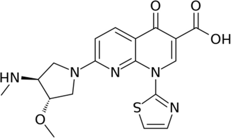
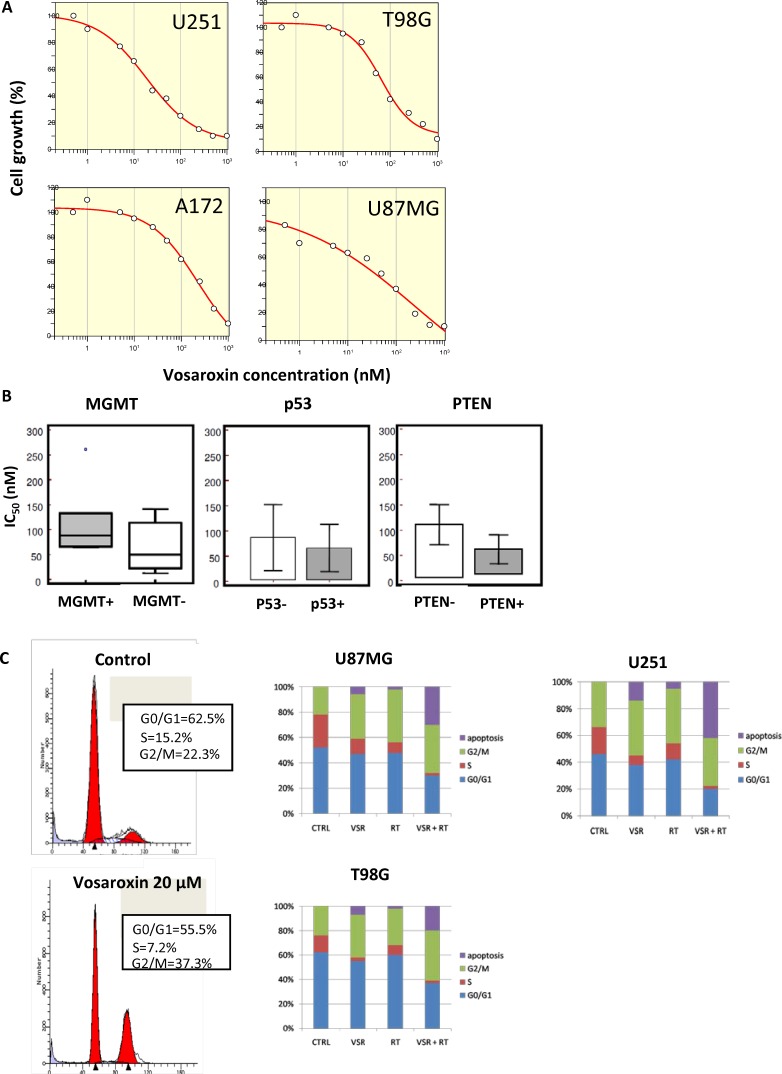
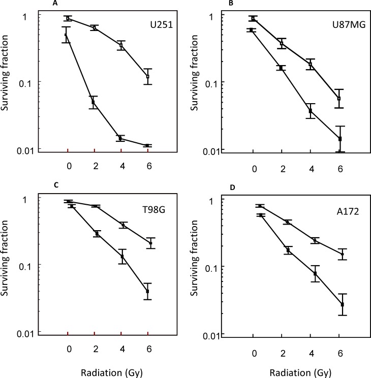
Purpose: Glioblastoma multiforme (GBM) is the most aggressive brain tumor. The activity of vosaroxin, a first-in-class anticancer quinolone derivative that intercalates DNA and inhibits topoisomerase II, was investigated in GBM preclinical models as a single agent and combined with radiotherapy (RT).

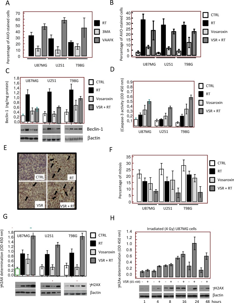
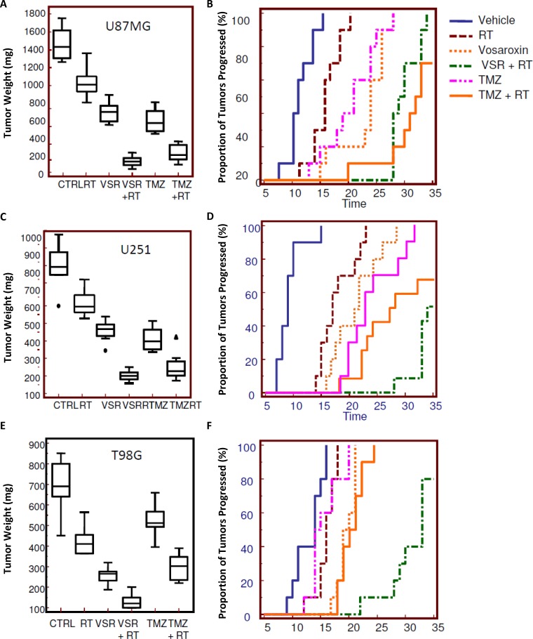
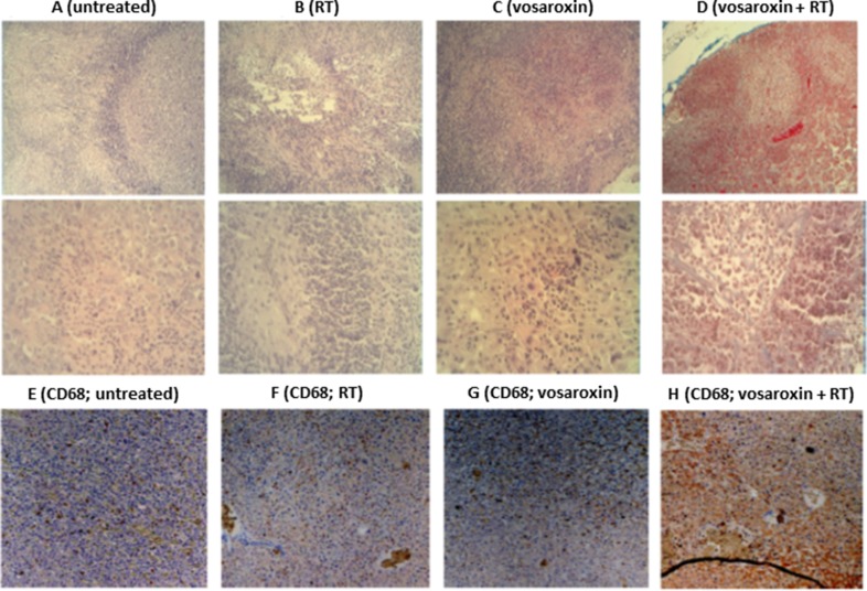
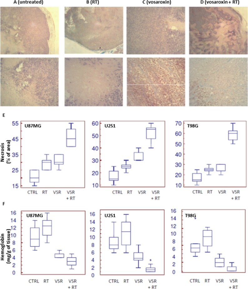
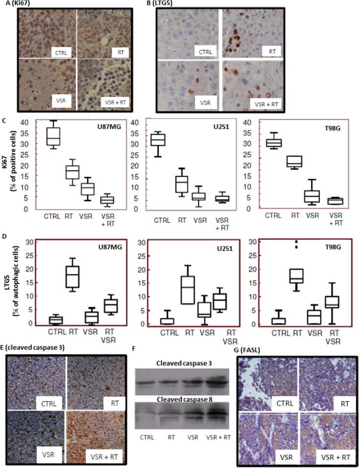
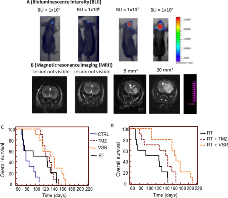
Results: Vosaroxin showed antitumor activity in clonogenic survival assays, with IC50 of 10-100 nM, and demonstrated radiosensitization. Combined treatments exhibited significantly higher γH2Ax levels compared with controls. In xenograft models, vosaroxin reduced tumor growth and showed enhanced activity with RT; vosaroxin/RT combined was more effective than temozolomide/RT. Vosaroxin/RT triggered rapid and massive cell death with characteristics of necrosis. A minor proportion of treated cells underwent caspase-dependent apoptosis, in agreement with in vitro results. Vosaroxin/RT inhibited RT-induced autophagy, increasing necrosis. This was associated with increased recruitment of granulocytes, monocytes, and undifferentiated bone marrow-derived lymphoid cells. Pharmacokinetic analyses revealed adequate blood-brain penetration of vosaroxin. Vosaroxin/RT increased disease-free survival (DFS) and overall survival (OS) significantly compared with RT, vosaroxin alone, temozolomide, and temozolomide/RT in the U251-luciferase orthotopic model.

Materials and methods: Cellular, molecular, and antiproliferative effects of vosaroxin alone or combined with RT were evaluated in 13 GBM cell lines. Tumor growth delay was determined in U87MG, U251, and T98G xenograft mouse models. (DFS) and (OS) were assessed in orthotopic intrabrain models using luciferase-transfected U251 cells by bioluminescence and magnetic resonance imaging.

Conclusions: Vosaroxin demonstrated significant activity in vitro and in vivo in GBM models, and showed additive/synergistic activity when combined with RT in O6-methylguanine methyltransferase-negative and -positive cell lines.

Keywords: double-strand breaks; glioblastoma; radiotherapy; topoisomerase II; vosaroxin.


Conflict of interest statement

CONFLICTS OF INTEREST

JAF was previously employed by and is a consultant for Sunesis Pharmaceuticals. GLG, AM, CM, FV, FM, AC, GR, LV, AV, EDC and CF have no conflicts of interest to disclose.

Figures

Figure 1
Figure 1. Chemical structure of vosaroxin
Figure 2
Figure 2. Effects of vosaroxin on glioma cell lines
(A) Growth inhibition curves for U251, T98G, A172, and U87MG glioma cell lines treated with vosaroxin, generated with Grafit software. (B) No statistically significant differences were found in the IC50 of vosaroxin in glioma cell lines by MGMT, p53, or PTEN status. (C) Cell cycle analysis of vosaroxin-induced G2/M cell cycle arrest (left panels), and combination effects of vosaroxin (20 nM) and radiotherapy on cell cycle arrest and apoptosis in U87MG, U251 and T98G cells (bar graphs in right panels).
Figure 3
Figure 3. Radiosensitizing effects of vosaroxin on glioma cell lines
Clonogenic curves for (A) U251, (B) U87MG, (C) T98G, and (D) A172 cells treated with vosaroxin (solid line) and control (no vosaroxin; dashed line).
Figure 4
Figure 4. Molecular and cytologic analyses of the mechanisms of radiosensitization by vosaroxin. To define the molecular mechanisms involved in radiosensitization by vosaroxin we treated cells with radiotherapy (RT; at 2, 4, and 6 Gy) and vosaroxin at concentrations equal to IC20 values (15 nM for U251, 65 nM for U87MG, and 45 nM for T98G cells)
(A) Percentage of acidic vesicular organelle (AVO)-stained glioma cells 24 hours after RT treatment (4 Gy). Percentage of AVO-stained cells was increased by the pan-caspase inhibitor z-Val-Ala-Asp(Ome)-fluoromethyl ketone (VAAFK, 10 μM) and was reduced by the autophagy inhibitor 3-methyladenine (3MA, 5 μM). (B) Percentage of AVO-stained cells after treatment with RT, vosaroxin (VSR), or the combination. CTRL: control. (C) Modulation of expression of an autophagy marker, beclin-1, by RT, vosaroxin, and combination vosaroxin plus RT by Western blot and ELISA. (D) Caspase-3 activity in treated and control cell cultures. (E) Immunocytochemical appearance of γH2Ax expression in T98G cultures after RT, vosaroxin, and combination RT plus vosaroxin for 24 hours. (F) Percentage of mitosis in U87MG, U251, and T98G cultures after single-agent or combination treatment. (G) γH2Ax expression in U87MG, U251, and T98G cells at 24 hours of treatment. (H) Expression of γH2Ax by ELISA and Western blotting measured in U87MG cells at 1, 4, 8, 16, 24, and 48 hours after 4 Gy irradiation with or without vosaroxin pre-treatment.
Figure 5
Figure 5. Radiosensitizing effects of vosaroxin on tumor weight and time to progression in xenograft models
To assess the effect on tumors in an in vivo model, 1 × 10⁶ cells of U251, U87MG, and T98G GBM cells were subcutaneously injected in female cd1 nu/nu mice. When tumors reached a volume of 80 mm3 (about 10 days after cell injection), animals were randomized to receive radiotherapy (RT) alone (1 single dose of 4 Gy), vosaroxin (VSR; 10 mg/kg q 5 d for 5 wk), or vosaroxin (10 mg/kg q 5 d for 5 wk) plus RT (1 single dose of 4 Gy administered after 3 days of vosaroxin treatment). These treatments were compared with standard therapies consisting of temozolomide (TMZ; 16 mg/kg ′ 5 consecutive days) and temozolomide plus RT. Changes in tumor volumes were measured over time. After 35 days, animals were sacrificed and tumors harvested and weighed. Final tumor weights (at day 35) and Kaplan-Meier analysis of time to progression are shown for: (A, B) U87MG; (C, D) U251; and (E, F) T98G xenograft models. CTRL: control.
Figure 6
Figure 6. Leukocyte infiltration with vosaroxin and/or RT
Panels (AD) show the histologic appearance of U251 xenografts at low (50′; upper row) and high (400′; lower row) magnification in (A) untreated U251 xenografts, (B) after radiotherapy (RT), (C) after treatment with vosaroxin, and (D) after vosaroxin was added to RT. Panels (EH) display immunostaining for CD68 expression at low magnification (50×) for untreated T98G tumors (E), and T98G tumors treated with RT (F), vosaroxin (G), and vosaroxin and RT (H).
Figure 7
Figure 7. Necrosis in U251 xenograft tumors treated with vosaroxin and/or RT
(AD) staining for necrosis in U251 xenografts at low (50×; upper row) and high (400×; lower row) magnification in untreated tumors (A) and tumors treated with RT (B), vosaroxin (C), and RT plus vosaroxin (D). (E) Graphical analysis on percentage of necrotic cells in U87MG, U251, and T98G xenografts after various treatments. (F) Quantification of the amount of hemoglobin (as an indirect measure of vasculature) present in tissue extracts from U87MG, U251, and T98G xenografts after various treatments. CTRL: control; RT: radiotherapy; VSR: vosaroxin.
Figure 8
Figure 8. Expression of proliferation, autophagic, and apoptotic markers
Immunohistochemical staining for expression of (A) Ki67 and (B) LTG5 in untreated and treated T98G xenografts. Quantification of Ki67 expression (C) and LTG5 expression (D) in U87MG, U251, and T98G xenografts. Immunohistochemical staining for caspase-3 expression (E) and Western blot analyses of caspase-3 and caspase-8 levels in tissue extracts (F) for untreated and treated T98G xenografts. (G) Immunohistochemical staining for FasL expression in the T98G xenograft model. CTRL: control; RT: radiotherapy; VSR: vosaroxin.
Figure 9
Figure 9. In vivo experiments: orthotopic intrabrain model
(A) Representative images of relative bioluminescence intensity (BLI) in brain lesion recurrence after orthotopic cell injections. (B) Representative MRI images of brain lesion recurrence. (C) Comparison of the effects of single treatments versus control (CTRL) on overall survival. (D) Comparison of the radiosensitizing effects of temozolomide (TMZ) and vosaroxin (VSR) on overall survival. Note: images in 9A and 9B are not representation of dose- or time-response data.

Similar articles

Cited by

References

    1. Polley MY, Lamborn KR, Chang SM, Butowski N, Clarke JL, Prados M. Six-month progression-free survival as an alternative primary efficacy endpoint to overall survival in newly diagnosed glioblastoma patients receiving temozolomide. Neuro Oncol. 2010;12:274–82. doi: 10.1093/neuonc/nop034. - DOI - PMC - PubMed
    1. Ringel F, Pape H, Sabel M, Krex D, Bock HC, Misch M, Weyerbrock A, Westermaier T, Senft C, Schucht P, Meyer B, Simon M, group SNs Clinical benefit from resection of recurrent glioblastomas: results of a multicenter study including 503 patients with recurrent glioblastomas undergoing surgical resection. Neuro Oncol. 2016;18:96–104. doi: 10.1093/neuonc/nov145. - DOI - PMC - PubMed
    1. Seidlitz A, Siepmann T, Lock S, Juratli T, Baumann M, Krause M. Impact of waiting time after surgery and overall time of postoperative radiochemotherapy on treatment outcome in glioblastoma multiforme. Radiat Oncol. 2015;10:172. doi: 10.1186/s13014-015-0478-5. - DOI - PMC - PubMed
    1. Kesari S. Understanding glioblastoma tumor biology: the potential to improve current diagnosis and treatments. Semin Oncol. 2011;38:S2–10. doi: 10.1053/j.seminoncol.2011.09.005. - DOI - PubMed
    1. Womeldorff M, Gillespie D, Jensen RL. Hypoxia-inducible factor-1 and associated upstream and downstream proteins in the pathophysiology and management of glioblastoma. Neurosurg Focus. 2014;37:E8. doi: 10.3171/2014.9.FOCUS14496. - DOI - PubMed

MeSH terms