. 2017 May 2;25(5):739-749.e3.
doi: 10.1016/j.str.2017.03.009. Epub 2017 Apr 13.

Flexibility and Design: Conformational Heterogeneity along the Evolutionary Trajectory of a Redesigned Ubiquitin

Affiliations

Flexibility and Design: Conformational Heterogeneity along the Evolutionary Trajectory of a Redesigned Ubiquitin

Justin T Biel et al. Structure. .

Abstract

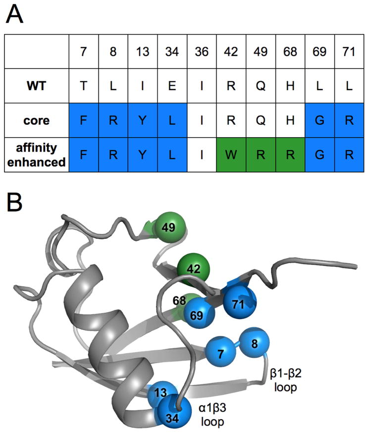
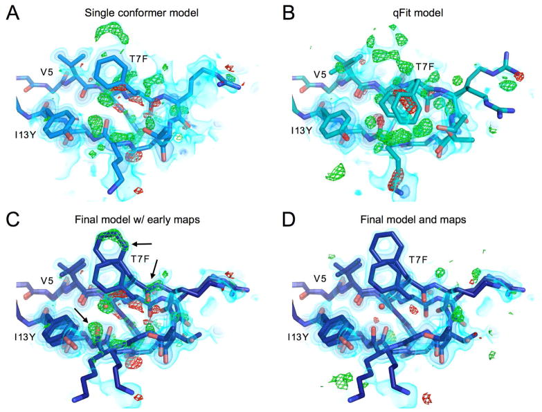
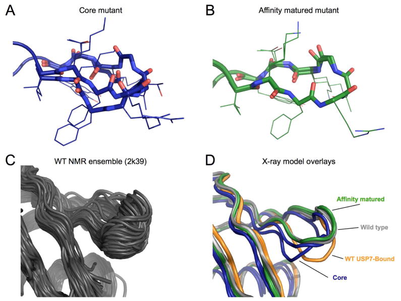
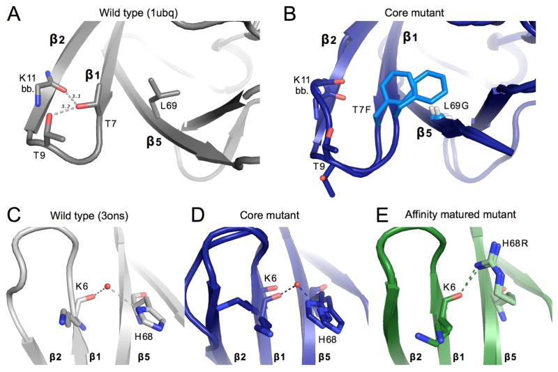
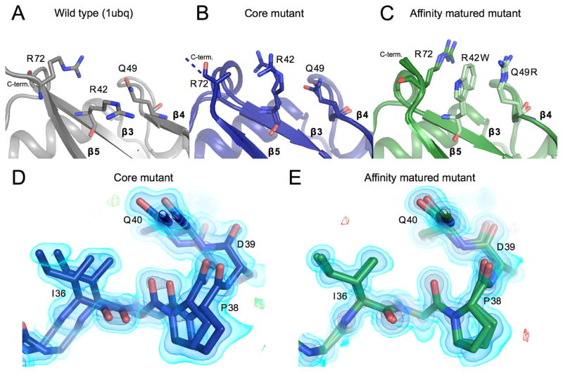
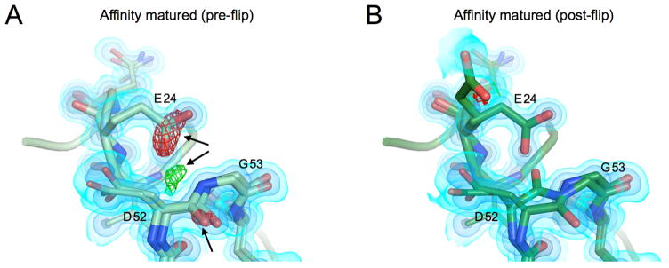
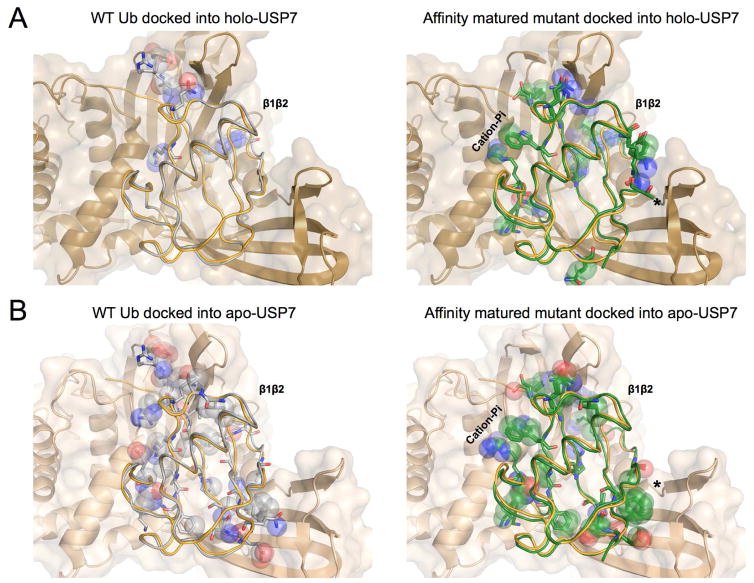
Although protein design has been used to introduce new functions, designed variants generally only function as well as natural proteins after rounds of laboratory evolution. One possibility for this pattern is that designed mutants frequently sample nonfunctional conformations. To test this idea, we exploited advances in multiconformer modeling of room-temperature X-ray data collection on redesigned ubiquitin variants selected for increasing binding affinity to the deubiquitinase USP7. Initial core mutations disrupt natural packing and lead to increased flexibility. Additional, experimentally selected mutations quenched conformational heterogeneity through new stabilizing interactions. Stabilizing interactions, such as cation-pi stacking and ordered waters, which are not included in standard protein design energy functions, can create specific interactions that have long-range effects on flexibility across the protein. Our results suggest that increasing flexibility may be a useful strategy to escape local minima during initial directed evolution and protein design steps when creating new functions.

Keywords: directed evolution; high-resolution X-ray crystallography; protein design; room-temperature X-ray crystallography; ubiquitin.


Figures

Figure 1
Figure 1. Locations and identities of mutations made across directed evolution trajectory of ubiquitin
A) Table showing amino acid identities of mutation sites for the wild-type, core and affinity matured mutants. Blue coloring represents residue identities first introduced in the core mutant. Green shows new residue identities for the affinity matured mutations. B) Wild-type ubiquitin (PDB ID: 1ubq) model shown in grey with spheres representing locations of mutation sites, colored as in panel A and labeled by residue number.
Figure 2
Figure 2
β1β2 loop of the core mutant showing residues 5–13, models and maps at different points in the refinement procedure A) Best single conformer model and corresponding 2mFo-DFc map shown in volume representation at three contour levels: 0.65 eÅ−3, 1.5 eÅ−3 and 3.5 eÅ−3 from lightest to darkest. mFo-DFc map shown in green and red mesh at 3 or −3 eÅ−3 respectively. B) Output model from qFit 2.0 built from single conformer model. While qFit is able to accurately model the side chain heterogeneity at the more ordered base of chain for residue 5 valine, qFit was unable to capture the backbone heterogeneity in this loop, and in fact may be mislead by the complex density as can be seen for the clearly misplaced Phe7 side chains. C) Final, manually-built multiconformer model with final 2mFo-DFc map, and single conformer difference map (mFo-DFc). This shows how the newly built heterogeneity corresponds to the major difference peaks in the original map. D) The final manually-built model with corresponding maps. While some difference features still exist, the difference signal in this region has largely been reduced in comparison to the original single conformer structure. See also Figure S1.
Figure 3
Figure 3. Conformations of the β1β2 loop
A) Final multiconformer model of the β1β2 loop for the core mutant. Backbone atoms in the loop are rendered in sticks, while the side chains are left as lines. B) Final model of β1β2 loop for the affinity matured mutant. Panel shown with electron density in Supplementary Figure 2. C) Backbone conformational ensemble from NMR relaxation dispersion experiments (PDB ID: 2k39). The majority of conformations exhibit a simple backbone shift, producing a hinge like motion as observed in previous MD simulations. D) β1β2 loop conformations from different ubiquitin structures. Wild-type ubiquitin apo-structure (PDB ID: 1ubq) in grey, and bound to USP7 in orange (PDB ID: 1nbf). The core mutant is shown in dark blue, and the affinity matured in green. See also Figure S2.
Figure 4
Figure 4. Structural changes upon mutation near β1β2-loop
A) Key packing and hydrogen bond interactions around mutation sites T7F and L69G. WT ubiquitin (1ubq) is shown in gray. Gray dashed lines show hydrogen bonds existent in WT ubiquitin between residues 7, 9, and 11. Sticks are shown for the side chains of residues 7 and 9, as well as for the backbone of residue 11. B) Both conformations of the residues shown in panel A are shown for the core mutant. Mutated residues are colored in a lighter blue. The Cα of Gly69 is shown as a small sphere for clarity. C, D, E) Interactions between residue 6 of β-stand 1 and residue 68 of strand 5 for the wild-type ubiquitin (gray, panel C), of the core mutant (blue, panel D), and the affinity matured mutant (green, panel E). A modeled water appears linking the backbone of residue 6 with the histidine 68 side-chain in both the core mutant and WT (3ons). This interaction is directly replaced in the affinity matured mutant by a hydrogen bond between the new arginine side-chain and the backbone carbonyl of residue 6. See also Figure S3.
Figure 5
Figure 5
Reduced conformational heterogeneity from core mutant to affinity matured mutant A, B, C) The packing of residues 42, 49, and 72 are shown. Panels and colors show the WT, core and affinity matured mutants in gray, blue and green respectively. Once mutated, residues are shown in a lighter shade of the same color. Residue Arg72 could not be fully built in the core mutant model and thus is truncated at the Cβ. Residues mutated in the affinity matured protein now show new cation-pi interactions, both between residues 72 and 42, and between 42 and 49. D) Residues 36–39 are shown highlighting heterogeneity in the affinity matured mutant that spans residues 32–41. There is signal for two conformations that differ in this region by a shift of as much as 2.2 Å. 2FoFc map shown as a volume contoured to 3.5, 1.5 and 0.65 eÅ−3 (light blue, blue, black), with the difference FoFc map contoured to 3 eÅ−3. E) The heterogeneity seen in the core mutant in panel D is not seen for the affinity matured structure at the same residues. Maps are contoured as in D. See also Figure S4.
Figure 6
Figure 6. Asp52-Gly53 peptide flip is seen in both states
A) Model and maps from the affinity matured mutation prior to modeling a peptide flip in this region. 2FoFc map shown as a volume contoured to 3.5, 1.5 and 0.65 eÅ−3 (light blue, blue, black), with the difference FoFc map contoured to 3 eÅ−3. There are clear difference features both positive (green) and negative (red) highlighted by black arrows. B) Modeled peptide flip in final structure of the core mutant. Maps are contoured as in A. The difference features are now gone.
Figure 7
Figure 7. WT and Affinity matured Ubiquitin have distinct patterns of contacts to Apo and holo USP7
A) Unbound WT Ub (grey - 1ubq) shown in ribbon overlaid with the bound Ub from the holo USP7 structure (orange - 1nbf). Ub-bound USP7 is shown in cartoon and surface (tan - 1nbf). Clashing atoms between the Unbound WT Ub and USP7 are mainly concentrated at the β1β2 loop (top) and shown in spheres with the remainder of the residue shown in sticks. B) Affinity enhanced mutant (green) shown overlaid with bound Ub from the holo USP7 structure. Although contacts are changed at the β1β2 loop (top), additional clashes, indicative of an altered binding mode or receptor accommodation are spread throughout the protein. An asterisk marks notable changes in clashes between apo and holo-USP7 structures (Panels B & D). C) Unbound WT Ub (grey -1ubq) shown overlaid with the unbound apo-USP7 structure (lighter brown - 5j7t). Overlay was constructed by alignment of domains to the bound-USP7 (1nbf). Increased clashes throughout the protein show the accommodation of the receptor in the holo form. D) Affinity matured mutant (green) shown overlaid with the unbound apo-USP7 structure (5j7t). Although clashes are increased in some regions, they are reduced in others, which suggests that the conformational flexibility of the receptor may be exploited by the Affinity matured mutant in the final binding pose. An asterisk marks notable changes in clashes between apo and holo-USP7 structures (Panels B & D).

Comment in

References

    1. Adams PD, Grosse-Kunstleve RW, Hung LW, Ioerger TR, McCoy AJ, Moriarty NW, Read RJ, Sacchettini JC, Sauter NK, Terwilliger TC. PHENIX: building new software for automated crystallographic structure determination. Acta Crystallogr D Biol Crystallogr. 2002;58:1948–1954. - PubMed
    1. Adhikary R, Yu W, Oda M, Walker RC, Chen T, Stanfield RL, Wilson IA, Zimmermann J, Romesberg FE. Adaptive mutations alter antibody structure and dynamics during affinity maturation. Biochemistry. 2015;54:2085–2093. - PMC - PubMed
    1. Bhabha G, Biel JT, Fraser JS. Keep on moving: discovering and perturbing the conformational dynamics of enzymes. Acc Chem Res. 2015;48:423–430. - PMC - PubMed
    1. Blomberg R, Kries H, Pinkas DM, Mittl PRE, Grütter MG, Privett HK, Mayo SL, Hilvert D. Precision is essential for efficient catalysis in an evolved Kemp eliminase. Nature. 2013;503:418–421. - PubMed
    1. Brünger AT, Adams PD, Rice LM. New applications of simulated annealing in X-ray crystallography and solution NMR. Structure. 1997;5:325–336. - PubMed

Publication types