

Astrocytes modulate thalamic sensory processing via mGlu2 receptor activation
- PMID: 28416443
- PMCID: PMC5480778
- DOI: 10.1016/j.neuropharm.2017.04.019
Astrocytes modulate thalamic sensory processing via mGlu2 receptor activation
Abstract
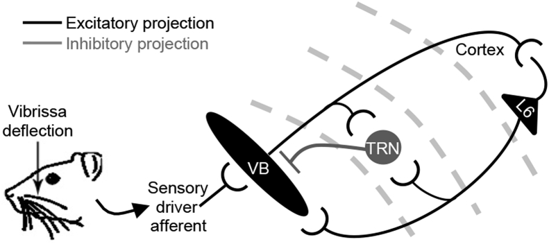
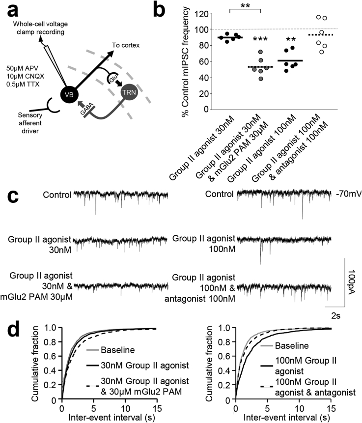
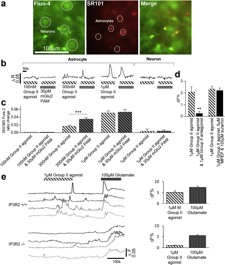
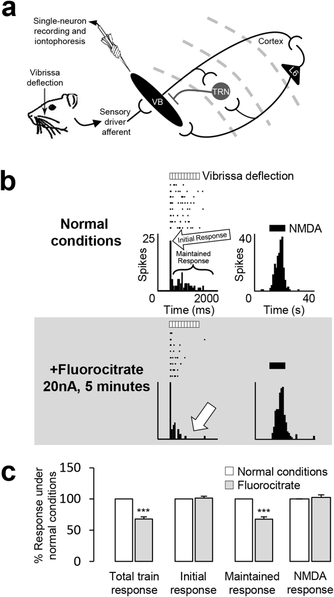
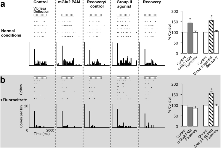
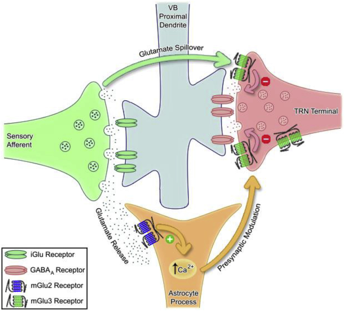
Astrocytes possess many of the same signalling molecules as neurons. However, the role of astrocytes in information processing, if any, is unknown. Using electrophysiological and imaging methods, we report the first evidence that astrocytes modulate neuronal sensory inhibition in the rodent thalamus. We found that mGlu2 receptor activity reduces inhibitory transmission from the thalamic reticular nucleus to the somatosensory ventrobasal thalamus (VB): mIPSC frequencies in VB slices were reduced by the Group II mGlu receptor agonist LY354740, an effect potentiated by mGlu2 positive allosteric modulator (PAM) LY487379 co-application (30 nM LY354740: 10.0 ± 1.6% reduction; 30 nM LY354740 & 30 μM LY487379: 34.6 ± 5.2% reduction). We then showed activation of mGlu2 receptors on astrocytes: astrocytic intracellular calcium levels were elevated by the Group II agonist, which were further potentiated upon mGlu2 PAM co-application (300 nM LY354740: ratio amplitude 0.016 ± 0.002; 300 nM LY354740 & 30 μM LY487379: ratio amplitude 0.035 ± 0.003). We then demonstrated mGlu2-dependent astrocytic disinhibition of VB neurons in vivo: VB neuronal responses to vibrissae stimulation trains were disinhibited by the Group II agonist and the mGlu2 PAM (LY354740: 156 ± 12% of control; LY487379: 144 ± 10% of control). Presence of the glial inhibitor fluorocitrate abolished the mGlu2 PAM effect (91 ± 5% of control), suggesting the mGlu2 component to the Group II effect can be attributed to activation of mGlu2 receptors localised on astrocytic processes within the VB. Gating of thalamocortical function via astrocyte activation represents a novel sensory processing mechanism. As this thalamocortical circuitry is important in discriminative processes, this demonstrates the importance of astrocytes in synaptic processes underlying attention and cognition.
Keywords: Astrocyte; Metabotropic glutamate receptor subtype 2; Synaptic inhibition; Thalamic reticular nucleus; Thalamus.
Copyright © 2017 The Authors. Published by Elsevier Ltd.. All rights reserved.
Figures
Similar articles
-
Positive allosteric modulation reveals a specific role for mGlu2 receptors in sensory processing in the thalamus.J Physiol. 2012 Feb 15;590(4):937-51. doi: 10.1113/jphysiol.2011.218065. Epub 2011 Dec 23. J Physiol. 2012. PMID: 22199165 Free PMC article.
-
Group II metabotropic glutamate receptor (mGlu2 and mGlu3 ) roles in thalamic processing.Br J Pharmacol. 2022 Apr;179(8):1607-1619. doi: 10.1111/bph.15640. Epub 2021 Sep 15. Br J Pharmacol. 2022. PMID: 34355803
-
Neuronal activity patterns in the mediodorsal thalamus and related cognitive circuits are modulated by metabotropic glutamate receptors.Neuropharmacology. 2015 May;92:16-24. doi: 10.1016/j.neuropharm.2014.12.031. Epub 2015 Jan 7. Neuropharmacology. 2015. PMID: 25576798 Free PMC article.
-
Pharmacological and molecular characterization of the positive allosteric modulators of metabotropic glutamate receptor 2.Neuropharmacology. 2016 Dec;111:253-265. doi: 10.1016/j.neuropharm.2016.08.032. Epub 2016 Aug 30. Neuropharmacology. 2016. PMID: 27590915 Review.
-
Reprint of Pharmacological and molecular characterization of the positive allosteric modulators of metabotropic glutamate receptor 2.Neuropharmacology. 2017 Mar 15;115:115-127. doi: 10.1016/j.neuropharm.2016.08.040. Epub 2017 Feb 16. Neuropharmacology. 2017. PMID: 28216000 Review.
Cited by
-
Group II Metabotropic Glutamate Receptors: Role in Pain Mechanisms and Pain Modulation.Front Mol Neurosci. 2018 Oct 9;11:383. doi: 10.3389/fnmol.2018.00383. eCollection 2018. Front Mol Neurosci. 2018. PMID: 30356691 Free PMC article. Review.
-
Rethinking Sensory Information Processing: The Essential Role of Astrocytes.J Neurochem. 2025 Jun;169(6):e70113. doi: 10.1111/jnc.70113. J Neurochem. 2025. PMID: 40490971 Free PMC article. Review.
-
Information Encoding in Bursting Spiking Neural Network Modulated by Astrocytes.Entropy (Basel). 2023 May 1;25(5):745. doi: 10.3390/e25050745. Entropy (Basel). 2023. PMID: 37238500 Free PMC article.
-
Enhanced mGluR5 intracellular activity causes psychiatric alterations in Niemann Pick type C disease.Cell Death Dis. 2024 Oct 23;15(10):771. doi: 10.1038/s41419-024-07158-8. Cell Death Dis. 2024. PMID: 39443481 Free PMC article.
-
Toll-Like Receptor 4 Knockdown Attenuates Brain Damage and Neuroinflammation After Traumatic Brain Injury via Inhibiting Neuronal Autophagy and Astrocyte Activation.Cell Mol Neurobiol. 2018 Jul;38(5):1009-1019. doi: 10.1007/s10571-017-0570-5. Epub 2017 Dec 8. Cell Mol Neurobiol. 2018. PMID: 29222622 Free PMC article.
References
-
- Alexander G.M., Godwin D.W. Unique presynaptic and postsynaptic roles of Group II metabotropic glutamate receptors in the modulation of thalamic network activity. Neuroscience. 2006;141:501–513. - PubMed
MeSH terms
Substances
LinkOut - more resources
Full Text Sources
Other Literature Sources
Research Materials
Miscellaneous
