. 2017 Jun 1;77(11):2881-2892.
doi: 10.1158/0008-5472.CAN-16-2204. Epub 2017 Apr 17.

NRF2 Induction Supporting Breast Cancer Cell Survival Is Enabled by Oxidative Stress-Induced DPP3-KEAP1 Interaction

Affiliations

NRF2 Induction Supporting Breast Cancer Cell Survival Is Enabled by Oxidative Stress-Induced DPP3-KEAP1 Interaction

Kevin Lu et al. Cancer Res. .

Abstract

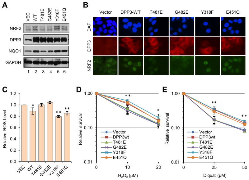
NRF2 is a transcription factor serving as a master regulator of the expression of many genes involved in cellular responses to oxidative and other stresses. In the absence of stress, NRF2 is constantly synthesized but maintained at low levels as it is targeted by KEAP1 for ubiquitination and proteasome-mediated degradation. NRF2 binds KEAP1 mainly through a conserved "ETGE" motif that has also been found in several other proteins, such as DPP3, which has been shown to bind KEAP1 and enhance NRF2 function upon overexpression. Here we demonstrate the interaction between endogenous DPP3 and endogenous KEAP1. We further show that the DPP3-KEAP1 interaction is strongly induced by hydrogen peroxide and that DPP3 is required for timely NRF2 induction and nuclear accumulation in the estrogen receptor (ER)-positive MCF7 breast cancer cells. Moreover, we present evidence that the binding of DPP3 to KEAP1 stabilizes the latter. Finally, we show that DPP3 is overexpressed in breast cancer and that elevated levels of DPP3 mRNA correlate with increased NRF2 downstream gene expression and poor prognosis, particularly for ER-positive breast cancer. Our studies reveal novel insights into the regulation of NRF2 and identify DPP3 and an NRF2 transcriptional signature as potential biomarkers for breast cancer prognosis and treatment. Cancer Res; 77(11); 2881-92. ©2017 AACR.


Figures

Figure 1
Figure 1
DPP3 interacts with KEAP1 in an oxidative stress-inducible manner through a highly conserved KEAP1-binding ETGE motif. (A) Composition of KEAP1 complexes isolated under non-stress and stress conditions. HeLa S3 cells stably expressing KEAP1 with FLAG-HA double tags at the C-terminus were either untreated or treated with 10 Gy ionizing radiation (IR), 100 μM tert-butylhydroxyquinone (tBHQ), 200 μM hydrogen peroxide (H2O2), or 2 mM hydroxy urea (HU). Cells were collected 2.5 hr after IR or drug treatment, and KEAP1-containing complexes were purified from the cytoplasmic and nuclear extracts of the cells by tandem affinity purification. The “Mock” purification was carried out using HeLa S3 cells without ectopic KEAP1. (B) Alignment of amino acid sequences of the “ETGE” or “ETGE”-like motifs and their immediate surrounding regions in proteins identified in the KEAP1 complexes purified under the “untreated” condition. CYT, cytosol; NUC, nucleus. (C-D) Oxidative stress-inducible interaction between DPP3 and KEAP1. Endogenous DPP3 was immunoprecipitated (IPed) from whole cell lysates of MCF7 cells treated with indicated concentrations of H2O2 for 3 hr (C) or diquat for 24 hr (D). Proteins in the IPed materials were analyzed by western blotting. (E) Kinetics of stress-induced DPP3 binding to KEAP1. MCF7 cells were treated with 200 μM H2O2 for indicated time periods, and the complex formation between DPP3 and KEAP1 was analyzed as above. (F) Requirement of the ETGE motif of DPP3 for KEAP1 binding. The wt and mutant DPP3 proteins were IPed with anti-FLAG beads from lysates of MCF7 cells stably expressing them. The IPed DPP3 and co-IPed KEAP1 were detected by western blotting.
Figure 2
Figure 2
DPP3 overexpression promotes NRF2 nuclear accumulation and ROS resistance. (A) Levels of DPP3, NRF2 and NQO1 proteins in MCF7 cells stably overexpressing wt or mutant DPP3 proteins. GAPDH was used as a loading control. (B) Localization of overexpressed DPP3 and endogenous NRF2 in the MCF7 stable cell lines. Immunofluorescence was carried out using anti-HA and anti-NRF2 antibodies for DPP3 and NRF2, respectively. (C) ROS levels in the MCF7 stable cell lines. (D-E) Sensitivities of the MCF7 stable cell lines to H2O2 (D) and diquat (E). Cells were treated with indicated concentrations of H2O2 and diquat for 42 hr. Values presented are means from 2 independent experiments. Error bars represent standard deviations (SDs). Statistical significance was calculated by Student's t test comparing the values for the 2 ETGE mutants (T481E and G482E) with those of 3 ETGE-wt proteins (wt, Y318F and E451Q). *p<0.05; **p<0.01.
Figure 3
Figure 3
Overexpression of DPP3 enhances the stability of KEAP1. (A) Levels of NRF2, DPP3, KEAP1 and p62 in MCF7 cell lines overexpressing wt and mutant DPP3 proteins. β-Actin was used a loading control. (B-C) Stabilities of KEAP in the MCF7 cell lines harboring the empty vector or overexpressing wt DPP3. Cells were either untreated or treated with 50 μg/ml of cycloheximide for 2, 4 and 6 hr, and the proteins were analyzed by western blotting. The intensities of KEAP1 bands were quantified by the ImageJ software, normalized against those of GAPDH and plotted. B shows a set of representative western blots, and C shows means of the quantified results from 3 independent experiments. Error bars represent SDs. *p<0.05. (D) Effect of DPP3 depletion in on KEAP1 levels in the stable MCF7 cell lines. The cells were treated with a control siRNA or siRNAs targeting DPP3 coding sequence (CDS) or 3′-UTR, and the proteins were analyzed by western blotting.
Figure 4
Figure 4
Depletion of DPP3 impairs NRF2 induction and sensitizes cells to hydrogen peroxide. (A-B) Depletion of DPP3 abrogates initial induction of NRF2. MCF7 cells were treated with transfection reagent alone (no siRNA), control siRNAs (NSC1 or AllStars) or 3 different DPP3 siRNAs for 72 hr in duplicates. One set of cells were then treated with 200 μM H2O2 for 2 hr, and the other set was left untreated (control). Proteins were analyzed by western blotting (A) and post induction NRF2 amounts were quantified by Image J (B). Data presented are means and SDs from 3 independent experiments. Statistical significance was calculated with Student's t test comparing the 2 control treated cells with the 3 DPP3 siRNA treated cells. ***, p<0.001. (C-D) Partial loss of DPP3 delays NRF2 induction. MCF7 cells were treated with transfection reagent alone (no siRNA) or a pool of 2 different DPP3 siRNAs for 72 hr and then with H2O2 for indicated periods. The amount and localization of NRF2 were analyzed by immunofluorescence (C) and western blotting (D). (E) Depletion of DPP3 sensitizes MCF7 cells to H2O2. Cells were treated with control or DPP3 siRNAs for 72 hr, H2O2 was added to indicated concentrations, and cell viability was measured 24 hr later. Values presented are means and error bars SDs from 3 independent experiments. Statistical significance was calculated with Student's t test comparing the 2 control siRNA treated cells with the 3 DPP3 siRNA treated cells. *, p<0.05; **, p<0.01.
Figure 5
Figure 5
DPP3 is overexpressed human breast cancer and correlates with increased NRF2 target gene expression and poor prognosis. (A) Box-and-whisker plots indicating the median score (horizontal line), the interquartile range (IQR, box boundaries) and 1.5 times the IQR (whiskers) demonstrate significantly higher DPP3 mRNA expression in 94 human breast tumors compared to 94 matched adjacent normal tissue samples (p=1.84×10-40, paired t-test). (B-C) A Spearman rank correlation demonstrating that DPP3 mRNA expression is positively correlated with DNA copy number status in (B) 1,031 TCGA breast tumor samples (p=1.6×10-96; r=0.5871) and (C) 1,992 samples from the METABRIC cohort (p=2.8×10-77, r=0.4019). (D) A Spearman rank correlation demonstrating that DPP3 and KEAP1 expression are positively correlated (p=7.6×10⁻¹¹, r=0.2009) in the TCGA cohort. DPP3 expression is positively associated with NRF2 target gene expression (p=5.4×10⁻¹⁴, r=0.2314) despite a negative correlation with NRF2 mRNA expression (p=2.8×10-08, r= [-0.1719]). Tumors with mutations in DPP3, KEAP1, NRF2, FH and KRAS are indicated with vertical bars. (E) Similar results as in D were observed in the METABRIC cohort (n=1,992). Breast cancer samples in D and E are ranked based on DPP3 mRNA expression; high KEAP1, NRF2 and NRF2 target gene expression is shown in red while low expression is indicated in blue. (F-H) Kaplan-Meier plots comparing disease-specific survival in human breast tumors from the METABRIC cohort based on high (top quartile) versus low (bottom quartile) DPP3 expression in (F) all tumors, (G) ER+ tumors, or (H) ER- tumors. (I-K) Kaplan-Meier plots comparing disease-specific survival in human breast tumors from the METABRIC cohort based on high (top quartile) versus low (bottom quartile) NRF2 target gene expression in (I) all tumors, (J) ER+ tumors, or (K) ER- tumors.

Similar articles

Cited by

References

    1. Taguchi K, Motohashi H, Yamamoto M. Molecular mechanisms of the Keap1-Nrf2 pathway in stress response and cancer evolution. Genes Cells. 2011;16(2):123–40. - PubMed
    1. Ma Q. Role of nrf2 in oxidative stress and toxicity. Annu Rev Pharmacol Toxicol. 2013;53:401–26. - PMC - PubMed
    1. Malhotra D, Portales-Casamar E, Singh A, Srivastava S, Arenillas D, Happel C, et al. Global mapping of binding sites for Nrf2 identifies novel targets in cell survival response through ChIP-Seq profiling and network analysis. Nucleic acids research. 2010;38(17):5718–34. - PMC - PubMed
    1. Agyeman AS, Chaerkady R, Shaw PG, Davidson NE, Visvanathan K, Pandey A, et al. Transcriptomic and proteomic profiling of KEAP1 disrupted and sulforaphane-treated human breast epithelial cells reveals common expression profiles. Breast Cancer Res Treat. 2011 - PMC - PubMed
    1. Suzuki T, Yamamoto M. Molecular basis of the Keap1-Nrf2 system. Free radical biology & medicine. 2015;88(Pt B):93–100. - PubMed

Publication types

Substances