Comparative Study
. 2017 Jun 8;129(23):3100-3110.
doi: 10.1182/blood-2016-11-751636. Epub 2017 Apr 17.

Erythrocyte sialoglycoproteins engage Siglec-9 on neutrophils to suppress activation

Affiliations
Comparative Study

Erythrocyte sialoglycoproteins engage Siglec-9 on neutrophils to suppress activation

Anel Lizcano et al. Blood. .

Abstract

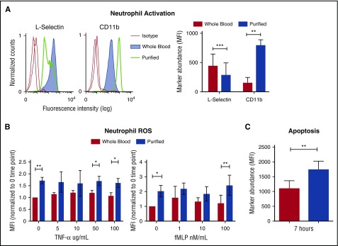
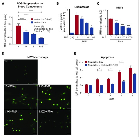
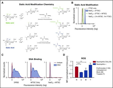
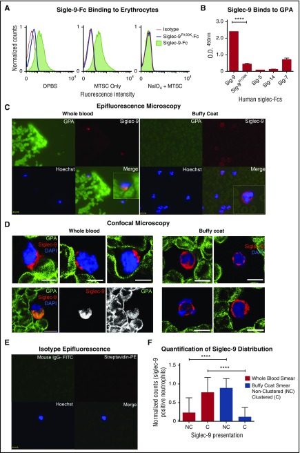
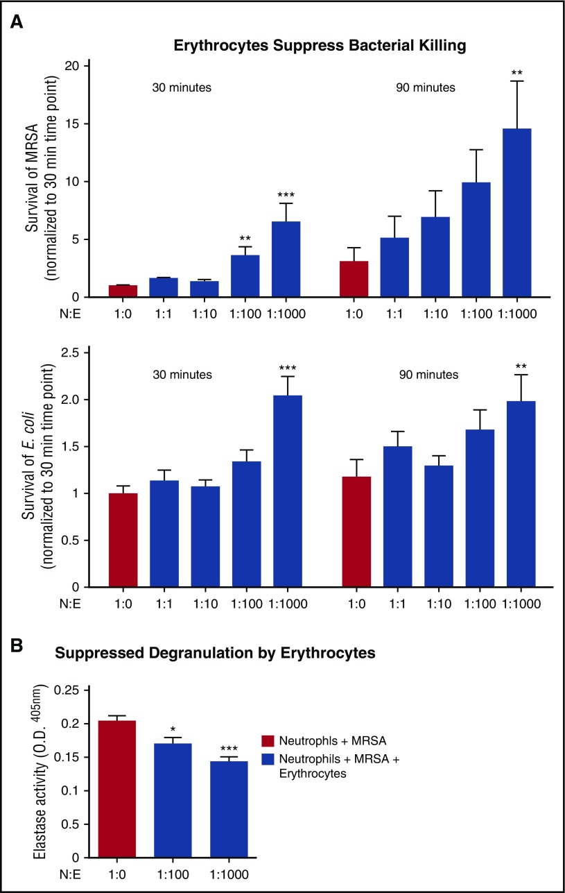
Healthy blood neutrophils are functionally quiescent in the bloodstream, have a short lifespan, and exit the circulation to carry out innate immune functions, or undergo rapid apoptosis and macrophage-mediated clearance to mitigate host tissue damage. Limitation of unnecessary intravascular neutrophil activation is also important to prevent serious inflammatory pathologies. Because neutrophils become easily activated after purification, we carried out ex vivo comparisons with neutrophils maintained in whole blood. We found a difference in activation state, with purified neutrophils showing signs of increased reactivity: shedding of l-selectin, CD11b upregulation, increased oxidative burst, and faster progression to apoptosis. We discovered that erythrocytes suppressed neutrophil activation ex vivo and in vitro, including reduced l-selectin shedding, oxidative burst, chemotaxis, neutrophil extracellular trap formation, bacterial killing, and induction of apoptosis. Selective and specific modification of sialic acid side chains on erythrocyte surfaces with mild sodium metaperiodate oxidation followed by aldehyde quenching with 4-methyl-3-thiosemicarbazide reduced neutrophil binding to erythrocytes and restored neutrophil activation. By enzyme-linked immunosorbent assay and immunofluorescence, we found that glycophorin A, the most abundant sialoglycoprotein on erythrocytes, engaged neutrophil Siglec-9, a sialic acid-recognizing receptor known to dampen innate immune cell activation. These studies demonstrate a previously unsuspected role for erythrocytes in suppressing neutrophils ex vivo and in vitro and help explain why neutrophils become easily activated after separation from whole blood. We propose that a sialic acid-based "self-associated molecular pattern" on erythrocytes also helps maintain neutrophil quiescence in the bloodstream. Our findings may be relevant to some prior experimental and clinical studies of neutrophils.


Figures

Figure 1.
Figure 1.
Purification and separation of human neutrophils from whole blood promotes neutrophil activation and apoptosis. (A) Heparinized human blood or purified neutrophils were evaluated for the expression of l-selectin and CD11b in resting conditions. Expression of cell surface markers was analyzed by flow cytometry and shows mean fluorescence intensity (MFI) gated on CD66b-positive cells. Histograms show the representative MFI from the 5 independent donors (left). Graph shows MFI, ± standard error of the mean (right). **P < .0066; ***P < .0004. (B) MFI of phagosomal ROS was analyzed from neutrophils in blood and purified neutrophils using Fc-OxyBURST Green assay reagent after 15 minutes and with the addition of TNF-α or fMLP; n = 4. *P < .0152 (left); *P < .0133 (right); **P < .0019. (C) Apoptosis of neutrophils was analyzed by TUNEL assay from neutrophils in whole blood and purified neutrophils at 7 hours. All CD66b+ neutrophils (3000 cell events) were analyzed for TUNEL MFI; n = 3. **P < .0020. All statistical data analyzed by Student paired t test versus control values. fMLP, formyl-methionyl-leucyl-phenylalanine; ROS, reactive oxygen species; TNF-α, tumor necrosis factor α; TUNEL, terminal deoxynucleotidyltransferase-mediated dUTP nick end labeling.
Figure 2.
Figure 2.
Autologous erythrocytes inhibit neutrophil oxidative burst, chemotaxis, extracellular trap formation and apoptosis. (A) Purified human neutrophils were coincubated in whole-blood components: plasma, erythrocytes, or both plasma and erythrocytes. Coincubation was done with whole plasma, and the erythrocyte concentration was at a 1:50 neutrophil:erythrocyte ratio. MFI of phagosomal ROS production was analyzed using Fc-OxyBURST Green assay reagent after 15 minutes; n = 3 Statistics were analyzed by ordinary 1-way analysis of variance (ANOVA). ****P < .0001. (B) The effect of neutrophil chemotaxis in combination with erythrocytes (neutrophils:erythrocytes; upper well) in the presence of 100 nM of fMLP (lower well) was determined using a transwell system. Statistics were analyzed by ordinary 1-way ANOVA; n = 4. *P < .03 versus control values considered statistically significant. (C) NET formation was quantified to determine the effect of erythrocytes incubated with neutrophils; n = 3. Statistics were analyzed by ordinary 1-way ANOVA. ***P < .0001 versus control values considered statistically significant. (D) NET formation microscopy analysis with increasing concentrations of erythrocytes plus phorbol myristate acetate (25 nM) where indicated. Scale bar, 50 μm. (E) Detection of apoptotic neutrophils was analyzed by TUNEL assay with and without incubation with erythrocytes up to 8 hours. Neutrophils were gated using forward and side scatter by flow cytometry and by TUNEL MFI of 10 000 cell counts. Results were analyzed by Student paired t test; n = 2. *P < .05 versus control values considered statistically significant. N:E, neutrophil:erythrocyte; PMA, phorbol myristate acetate; RFU, relative fluorescence units.
Figure 3.
Figure 3.
Modification of side chains of terminal sialic acids on erythrocyte surface by mild periodate and MTSC. (A) Mild oxidation using sodium periodate (NaIO4) generates aldehydes on sialic acid–containing glycoproteins, followed by direct labeling of aldehydes with a fluorescent tag, FTSC (top, green). A smaller compound, MTSC, replaced FTSC, which would generate the same sialic acid modification without the fluorescein molecule (bottom, blue). (B) By flow cytometry, modification of sialic acid by both FTSC and MTSC was tested for reactivity and competition on the erythrocytes surface. Erythrocytes were treated with sodium periodate (NaIO4) for 20 minutes on ice, followed by the addition of FTSC or MTSC, where noted, for 1 hour at 37°C; n = 2. (C) Sialic acid modification of treated erythrocytes (NaIO4 + MTSC, MTSC only) and untreated erythrocytes (DPBS) were stained with biotinylated SNA lectin, which preferentially binds to sialic acids attached to terminal galactose in α2-6-linkage and was measured by flow cytometry. Isotype control (streptavidin-PE); n = 4. (D) Purified neutrophils were incubated with erythrocytes (DPBS, MTSC only, and NaIO4 + MTSC). Erythrocyte concentration was at 1:50 neutrophil:erythrocyte ratio. MFI of phagosomal ROS production was analyzed using Fc-OxyBURST Green assay reagent at 15 minutes. Statistics were analyzed by ordinary 1-way ANOVA; n = 2. **P < .0072 versus control values considered statistically significant; ***P < .0004.
Figure 4.
Figure 4.
GPA, the major surface sialoglycoprotein on erythrocytes, engages Siglec-9 via sialic acids. (A) Treated (NaIO4/MTSC), mock treated (MTSC only), and untreated (DPBS) erythrocytes were evaluated for the binding of Siglec-9-Fc and analyzed by flow cytometry. Isotype control (goat anti-human IgG); n = 4. (B) Five human recombinant Siglec-Fcs were measured for their ability to bind immobilized GPA by enzyme-linked immunosorbent assay; n = 3. Statistics were analyzed by ordinary 1-way ANOVA. ****P < .0001 versus control values considered statistically significant. (C) Smears prepared immediately after drawing whole blood (left) or those prepared from buffy coat (right), fixed and costained for the erythrocyte GPA (green), Siglec-9 on neutrophils (red), and nucleus (Hoechst). Clustering (Merge) of GPA with neutrophils Siglec-9 upon contact (1 representative image of each shown). Scale bar, 10 μm. (D) Smears were stained similarly as in panel C and analyzed by confocal microscopy. Whole-blood smears (left) confirm polarized clustering of Siglec-9 on neutrophils toward GPA-covered red blood cells (4 representative patterns are shown). One staining pattern in which GPA colocalizes with Siglec-9 (lower left); individual GPA and Siglec-9 channels are in black and white. Scale bars, 5 µm. (E) Isotype controls were stained with streptavidin-PE, mouse IgG1-FITC, and Hoechst stain. Scale bar, 10 μm. (F) Siglec-9 clustering and nonclustering in neutrophils from epifluorescent blood smears were counted and normalized to the total number of Siglec-9 positive neutrophils. Representative images from panel C or pooled data percentage (normalized to Siglec-9 positive cells) are shown (+/− standard error of the mean). Statistical analysis was performed using ordinary 1-way ANOVA with Holm–Sidak's multiple comparison test. ****P < .0001. DAPI, 4′,6-diamidino-2-phenylindole; O.D., optical densitiy; Sig, Siglec.
Figure 5.
Figure 5.
Erythrocytes suppress neutrophils in whole blood, but do not completely suppress bacterial killing. (A) Neutrophil killing of MRSA or E coli K12 was evaluated with increasing concentrations of erythrocytes at 30 and 90 minutes. Neutrophil killing of MRSA (top). Positive control is neutrophils plus MRSA. Statistics were analyzed by repeated measures one-way ANOVA and post hoc Holm–Sidak's multiple comparison test; n = 3. **P < .0057; ***P < .0001 versus control values considered statistically significant. Neutrophil killing of E coli (bottom). Positive control is neutrophils plus E coli. Statistics were analyzed by repeated measures 1-way ANOVA and post hoc Holm–Sidak's multiple comparison test; n = 3. **P < .05 versus control values considered statistically significant; ***P < .0003. (B) Neutrophils were incubated with MRSA for 90 minutes, and degranulation was measured by elastase release on fluorescent plate reader at ex. 488/emm.530; n = 3. Statistics were analyzed by ordinary 1-way ANOVA. ***P < .0001 versus control values considered statistically significant. N:E, neutrophil:erythrocyte.

Similar articles

Cited by

References

    1. Mohandas N. Structure and composition of the erythrocyte. In: Kaushansky EA, ed. Williams Hematology. New York, NY: McGraw-Hill; 2016:461-478.
    1. Belcher JD, Chen C, Nguyen J, et al. Heme triggers TLR4 signaling leading to endothelial cell activation and vaso-occlusion in murine sickle cell disease. Blood. 2014;123(3):377-390. - PMC - PubMed
    1. Chen G, Zhang D, Fuchs TA, Manwani D, Wagner DD, Frenette PS. Heme-induced neutrophil extracellular traps contribute to the pathogenesis of sickle cell disease. Blood. 2014;123(24):3818-3827. - PMC - PubMed
    1. Silvestre-Roig C, Hidalgo A, Soehnlein O. Neutrophil heterogeneity: implications for homeostasis and pathogenesis. Blood. 2016;127(18):2173-2181. - PubMed
    1. Summers C, Rankin SM, Condliffe AM, Singh N, Peters AM, Chilvers ER. Neutrophil kinetics in health and disease. Trends Immunol. 2010;31(8):318-324. - PMC - PubMed

Publication types

MeSH terms