. 2017 Aug;76(8):1440-1448.
doi: 10.1136/annrheumdis-2016-210706. Epub 2017 Apr 17.

Semaphorin 4D inhibits neutrophil activation and is involved in the pathogenesis of neutrophil-mediated autoimmune vasculitis

Affiliations

Semaphorin 4D inhibits neutrophil activation and is involved in the pathogenesis of neutrophil-mediated autoimmune vasculitis

Masayuki Nishide et al. Ann Rheum Dis. 2017 Aug.

Abstract

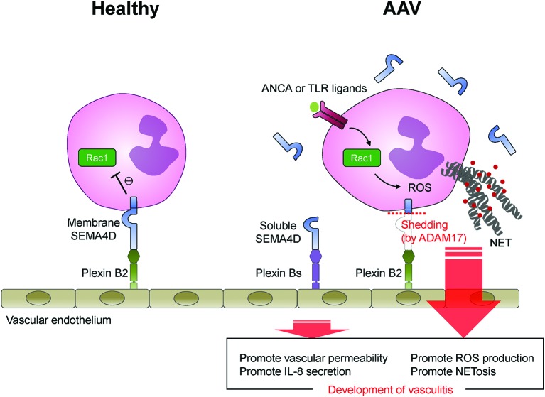
Objectives: Inappropriate activation of neutrophils plays a pathological role in antineutrophil cytoplasmic antibody (ANCA)-associated vasculitis (AAV). The aim of this study was to investigate the functions of semaphorin 4D (SEMA4D) in regulation of neutrophil activation, and its involvement in AAV pathogenesis.

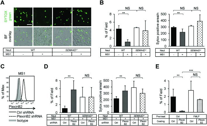
Methods: Serum levels of soluble SEMA4D were evaluated by ELISA. Blood cell-surface expression of membrane SEMA4D was evaluated by flow cytometry. To determine the functional interactions between neutrophil membrane SEMA4D and endothelial plexin B2, wild-type and SEMA4D-/- mice neutrophils were cultured with an endothelial cell line (MS1) stained with SYTOX green, and subjected to neutrophil extracellular trap (NET) formation assays. The efficacy of treating human neutrophils with recombinant plexin B2 was assessed by measuring the kinetic oxidative burst and NET formation assays.

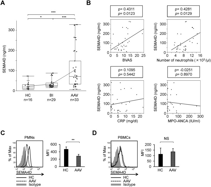
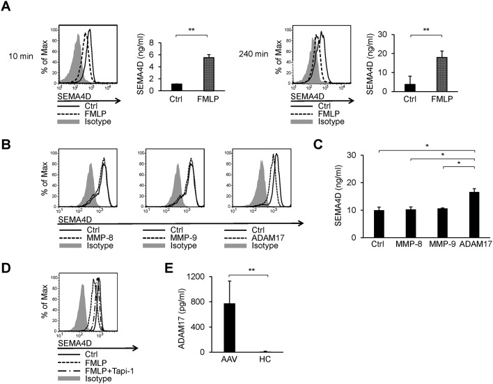
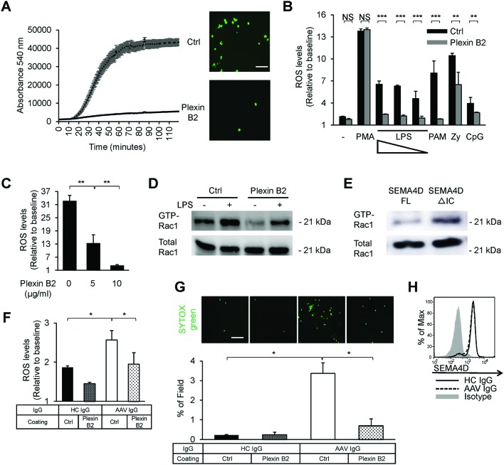
Results: Serum levels of soluble SEMA4D were elevated in patients with AAV and correlated with disease activity scores. Cell-surface expression of SEMA4D was downregulated in neutrophils from patients with AAV, a consequence of proteolytic cleavage of membrane SEMA4D. Soluble SEMA4D exerted pro-inflammatory effects on endothelial cells. Membranous SEMA4D on neutrophils bound to plexin B2 on endothelial cells, and this interaction decreased NET formation. Recombinant plexin B2 suppressed neutrophil Rac1 activation through SEMA4D's intracellular domain, and inhibited pathogen-induced or ANCA-induced oxidative burst and NET formation.

Conclusions: Neutrophil surface SEMA4D functions as a negative regulator of neutrophil activation. Proteolytic cleavage of SEMA4D as observed in patients with AAV may amplify neutrophil-mediated inflammatory responses. SEMA4D is a promising biomarker and potential therapeutic target for AAV.

Keywords: Autoimmune Diseases; Granulomatosis with polyangiitis; Inflammation; Systemic vasculitis.


Conflict of interest statement

Competing interests: None declared.

Figures

Figure 1
Figure 1
Identification of semaphorin 4D (SEMA4D) as a neutrophil activation marker associated with ANCA-associated vasculitis (AAV). (A) Soluble SEMA4D levels in serum samples from 16 healthy individuals (HC), 29 patients with bacterial infections (BI) and 33 patients with AAV. (B) Correlation of soluble SEMA4D serum levels with Birmingham Vasculitis Activity Score (BVAS) 2008 version 3, blood neutrophil counts, C reactive protein (CRP) levels and MPO-ANCA titres. Correlations were quantified using Spearman’s rank correlation coefficient (ρ). Flow cytometry of SEMA4D cell-surface expression by peripheral blood polymorphonuclear cells (PMNs) (C) and mononuclear cells (PBMCs) (D) from healthy individuals (HC; solid line) and patients with AAV (AAV; dotted line). Cells from HC were also stained with an isotype-matched control antibody (isotype; grey-filled histogram). Mean fluorescence intensity (MFI) profiles were determined from five HC or five patients with AAV. NS, not significant (p>0.05); *p<0.05; **p<0.01; ***p<0.001, as determined by a non-parametric Mann-Whitney U test (A) or a two-tailed unpaired Student’s t-test (C–D). Values are means±SD (A) and means±SEM (C–D).
Figure 2
Figure 2
Shedding of semaphorin 4D (SEMA4D) in stimulated neutrophils. (A) Cell-surface expression of SEMA4D on isolated human neutrophils derived from healthy donors, stimulated with or without formyl-methionyl-leucyl-phenylalanine (FMLP). Flow cytometry was performed at the indicated time points (10 or 240 min) on neutrophils cultured with control medium containing 2 µM FMLP (dotted line) or control medium (Ctrl; solid line). Cells from the control medium were also stained with an isotype-matched control antibody (isotype; grey-filled histogram). Soluble SEMA4D levels in the culture supernatants at each time point were determined by ELISA. (B) SEMA4D cell-surface expression on isolated human neutrophils stimulated with metalloproteases. Isolated neutrophils were primed with tumour necrosis factor (TNF)-α (2 ng/mL) for 30 min, and then stimulated either recombinant matrix metalloprotease (MMP)-8 (5 µg/mL), MMP-9 (5 µg/mL) or TNF-α converting enzyme (ADAM17; 5 µg/mL) for 60 min. Flow cytometry was performed similarly to (A). Soluble SEMA4D levels in each culture supernatant were determined by ELISA (C). (D) Effects of ADAM17 inhibitor Tapi-1 on cell-surface cleavage of SEMA4D. Isolated human neutrophils were pretreated with Tapi-1 (40 µM) for 30 min, followed by stimulation with FMLP (2 µM) for 30 min. Flow cytometry was performed similarly to (A). (E) Serum levels of ADAM17 in 33 patients with ANCA-associated vasculitis (AAV) and 16 healthy individuals (HC). NS, not significant (p>0.05); *p<0.05; **p<0.01; ***p<0.001, as determined by a two-tailed unpaired Student’s t-test (A, C) or a non-parametric Mann-Whitney U test (E). Values are means±SEM (A, C) and means±SD (E). Data are representative of three independent experiments (A–D).
Figure 3
Figure 3
Interaction between neutrophil semaphorin 4D (SEMA4D) and endothelial plexin B2 negatively regulates neutrophil extracellular trap (NET) formation. (A) SYTOX green immunofluorescence analysis was performed on bone marrow-derived neutrophils isolated from wild-type (WT) or SEMA4D-deficient (SEMA4D−/−) mice cocultured with or without MS1 cells and treated with lipopolysaccharide (LPS) (10 µg/mL) for 4 hours to induce NET formation. Scale bar: 100 µm. (B) Quantification of NET formation. Data are expressed as total SYTOX green-positive extracellular DNA area (μm2) per field (over 10 randomly chosen fields in each experiment) and average SYTOX green-positive area (μm2) was normalised against the total number of SYTOX-positive neutrophils in the same fields (400–500 neutrophils per field were counted and averaged in each experiment). (C) Knockdown of plexin B2 in MS1 cells. Cell-surface expression of plexin B2 was analysed using flow cytometry. MS1 cells treated with control short hairpin RNA (shRNA) were also stained with the isotype-matched control antibody (isotype; grey-filled histogram). (D) Effects of plexin B2 knockdown in MS1 cells on NET formation. Quantification of NET formation was performed similarly to (B). (E) Effects of plexin B2 on human NET formation. Isolated neutrophils derived from healthy donors were stimulated with or without 2 µM FMLP, washed and subsequently seeded onto culture plates precoated with recombinant human plexin B2 protein or control his-tag protein. These cells were incubated for 60 min, then stimulated with LPS (10 µg/mL) for 4 hours. SYTOX green immunofluorescence analysis was performed and evaluated similarly to (A and B). NS, not significant (p>0.05); *p<0.05; **p<0.01; ***p<0.001, as determined by a two-tailed unpaired Student’s t-test. Values are means±SEM (B, D, E). Data are representative of three independent experiments (A–E).
Figure 4
Figure 4
Inhibition of the neutrophil oxidative burst by the semaphorin 4D (SEMA4D)-plexin B2 interaction. (A) Effects of plexin B2 on the generation of reactive oxygen species (ROS) in human neutrophils. Neutrophils isolated from healthy donors were seeded onto culture plates precoated with recombinant human plexin B2 or control his-tag protein. These cells were incubated for 60 min, then stimulated with lipopolysaccharide (LPS) (1 µg/mL) for 2 hours. ROS levels were monitored every 1 min for a total of 120 cycles, and representative traces are the means from one experiment performed in triplicate. SYTOX green immunofluorescence analysis was performed at the last period of 4 hours after LPS stimulation, and image of neutrophil extracellular trap (NET) formation from each culture was attached. Scale bar: 100 µm. (B) Effects of plexin B2 on the generation of ROS in response to various stimulants. Isolated neutrophils were seeded similarly to (A) and were either left unstimulated (−) or stimulated with Phorbol 12-myristate 13-acetate (PMA) (1 µM), LPS (100 µg/mL, 1 µg/mL and 100 ng/mL), Pam3CSK4 (1 µg/mL), Zymosan (1 µg/mL) or CpG-ODN (1 µg/mL) for 2 hours. Relative ROS levels were determined as the ratio of fluorescent peak to baseline. (C) Dose-dependent effects of plexin B2 on ROS generation. Isolated neutrophils were seeded and stimulated similarly to (A), on culture plates precoated with recombinant human plexin B2 at the indicated concentrations. (D) Effects of plexin B2 on Rac1 activation in human neutrophils. Isolated neutrophils were seeded and incubated for 60 min similarly to (A) (LPS−), then stimulated with LPS (1 µg/mL) for 20 min (LPS+). Cells were lysed at each time point, and GTP-bound Rac1 was pulled down using GST-PAK-CRIB as described. Proteins were separated by SDS-PAGE (5×10⁶ cells/lane) and blotted with antihuman Rac1 antibody. (E) GTP-bound Rac1 pull-down assay using SEMA4D-transfected TLR4-expressing HEK293 cells. TLR4-expressing HEK293 cells were transfected with a construct expressing either full-length SEMA4D (FL) or SEMA4D without the intracellular C-terminal domain (ΔIC). Subsequently, cells were stimulated with LPS (1 µg/mL; 40 min) and lysed, then GTP-bound Rac1 was pulled down and blotted similarly to (D). Effects of plexin B2 on ROS production (F) and NET formation (G) in response to purified IgG from a healthy donor (HC IgG) or a patient with AAV (AAV IgG). Isolated neutrophils were primed with tumour necrosis factor (TNF)-α (2 ng/mL) for 15 min, then seeded similarly to (A), and stimulated with each immunoglobulin (200 µg/mL) for 1 hour (ROS) or 4 hours (NET). NET formation was evaluated similarly to (figure 3B). Scale bar: 200 µm. (H) Flow cytometry of cell-surface SEMA4D on neutrophils, primed and stimulated similarly to (F). NS, not significant (p>0.05); *p<0.05; **p<0.01; ***p<0.001, as determined by a two-tailed unpaired Student’s t-test. Values are means±SEM (A–C, F and G). Data are representative of three independent experiments (A–H).
Figure 5
Figure 5
Graphical scheme of this study. Under healthy conditions, the interaction between endothelial plexin B2 ligand and neutrophil cells surface SEMA4D receptor inhibits Rac1 activation and negatively regulates the generation of ROS and NET formation. By contrast, in patients with AAV, neutrophil surface SEMA4D is shed by ADAM17. Soluble SEMA4D had pro-inflammatory functions on endothelial cells. In addition, alteration of the SEMA4D-plexin B2 interaction results in aberrant activation of neutrophils, and this dichotomous effect is involved in the pathogenesis of AAV.

Similar articles

Cited by

References

    1. Nauseef WM, Borregaard N. Neutrophils at work. Nat Immunol 2014;15:602–11. 10.1038/ni.2921 - DOI - PubMed
    1. Phillipson M, Kubes P. The neutrophil in vascular inflammation. Nat Med 2011;17:1381–90. 10.1038/nm.2514 - DOI - PMC - PubMed
    1. Brinkmann V, Reichard U, Goosmann C, et al. . Neutrophil extracellular traps kill Bacteria. Science 2004;303:1532–5. 10.1126/science.1092385 - DOI - PubMed
    1. Brinkmann V, Zychlinsky A. Neutrophil extracellular traps: is immunity the second function of chromatin? J Cell Biol 2012;198:773–83. 10.1083/jcb.201203170 - DOI - PMC - PubMed
    1. Ji JY, Jing H, Diamond SL. Hemodynamic regulation of inflammation at the endothelial-neutrophil interface. Ann Biomed Eng 2008;36:586–95. 10.1007/s10439-008-9465-4 - DOI - PubMed

MeSH terms