

Potential Toxicity of Polymyxins in Human Lung Epithelial Cells
- PMID: 28416543
- PMCID: PMC5444173
- DOI: 10.1128/AAC.02690-16
Potential Toxicity of Polymyxins in Human Lung Epithelial Cells
Abstract
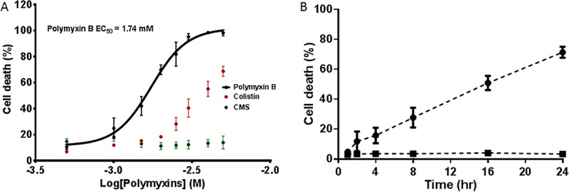
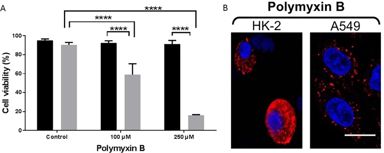
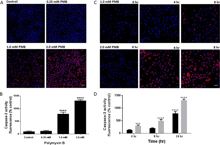
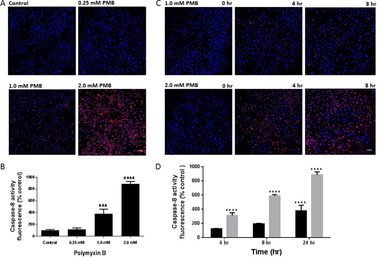
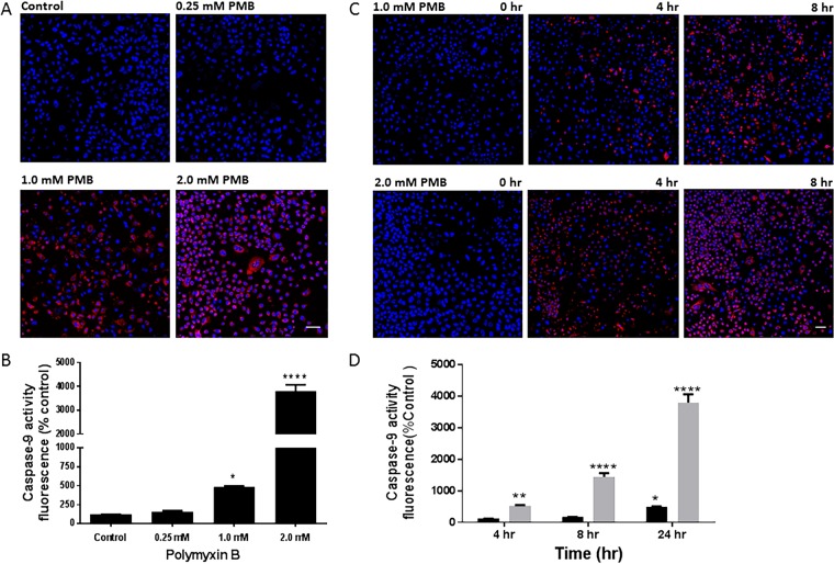
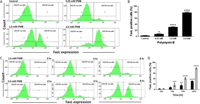
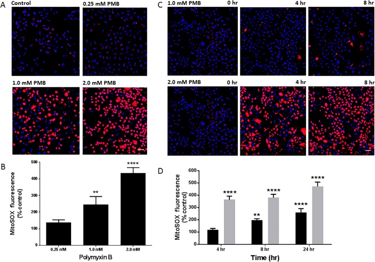
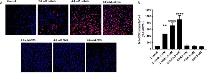
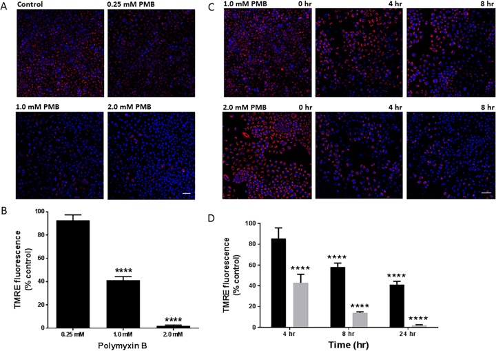
Inhaled polymyxins are of considerable utility in achieving optimal exposure in the respiratory tract for the treatment of lung infections caused by multidrug-resistant Gram-negative pathogens. Current inhaled polymyxin therapy is empirical, and often large doses are used that may lead to potential pulmonary adverse effects. This study aimed to investigate the effect of polymyxins on human lung epithelial (A549) cells. The viability of A549 cells was examined after treatment with polymyxins by flow cytometry. Activation of caspases 3, 8, and 9, expression of Fas ligand (FasL), loss of mitochondrial membrane potential, and mitochondrial oxidative stress induced by polymyxin B were evaluated. The concentration of polymyxin B required to induce 50% of maximal cell death was 1.74 mM (95% confidence interval, 1.60 to 1.90 mM). Colistin was at least 2-fold less toxic than polymyxin B, while colistimethate was nontoxic. With 2.0 mM polymyxin B, 30.6% ± 11.5% (mean ± standard deviation) of the cells were apoptotic at 8 h and this increased to 71.3% ± 3.72% at 24 h. Concentration- and time-dependent activation of caspases 3, 8, and 9 was evident, while the activation of caspase 9 was more dramatic. Furthermore, polymyxin B caused concentration- and time-dependent FasL expression, production of mitochondrial reactive oxygen species, and changes in mitochondrial membrane potential. This is the first study to demonstrate that both extrinsic death receptor and intrinsic mitochondrial pathways are involved in polymyxin-induced toxicity in A549 cells. This knowledge base is critical for the development of novel strategies for the safe and effective inhalation therapy of polymyxins against Gram-negative "superbugs."
Keywords: apoptosis; mitochondria; polymyxin; pulmonary delivery; respiratory toxicity.
Copyright © 2017 American Society for Microbiology.
Figures
Similar articles
-
Major pathways of polymyxin-induced apoptosis in rat kidney proximal tubular cells.Antimicrob Agents Chemother. 2015 Apr;59(4):2136-43. doi: 10.1128/AAC.04869-14. Epub 2015 Jan 26. Antimicrob Agents Chemother. 2015. PMID: 25624331 Free PMC article.
-
Polymyxin-Induced Metabolic Perturbations in Human Lung Epithelial Cells.Antimicrob Agents Chemother. 2021 Aug 17;65(9):e0083521. doi: 10.1128/AAC.00835-21. Epub 2021 Aug 17. Antimicrob Agents Chemother. 2021. PMID: 34228550 Free PMC article.
-
Pharmacokinetics/pharmacodynamics of colistin and polymyxin B: are we there yet?Int J Antimicrob Agents. 2016 Dec;48(6):592-597. doi: 10.1016/j.ijantimicag.2016.09.010. Epub 2016 Oct 18. Int J Antimicrob Agents. 2016. PMID: 27793510 Free PMC article. Review.
-
Framework for optimisation of the clinical use of colistin and polymyxin B: the Prato polymyxin consensus.Lancet Infect Dis. 2015 Feb;15(2):225-34. doi: 10.1016/S1473-3099(14)70850-3. Epub 2014 Oct 21. Lancet Infect Dis. 2015. PMID: 25459221
-
Polymyxin B, colistin, and sodium colistimethate.Med Clin North Am. 1982 Jan;66(1):135-42. doi: 10.1016/s0025-7125(16)31447-x. Med Clin North Am. 1982. PMID: 6278236 Review. No abstract available.
Cited by
-
Polymyxin B Peptide Hydrogel Coating: A Novel Approach to Prevent Ventilator-Associated Pneumonia.Int J Mol Sci. 2024 Sep 24;25(19):10269. doi: 10.3390/ijms251910269. Int J Mol Sci. 2024. PMID: 39408597 Free PMC article.
-
Effects of the antibiotic component on in-vitro bacterial killing, physico-chemical properties, aerosolization and dissolution of a ternary-combinational inhalation powder formulation of antibiotics for pan-drug resistant Gram-negative lung infections.Int J Pharm. 2019 Apr 20;561:102-113. doi: 10.1016/j.ijpharm.2019.02.018. Epub 2019 Feb 21. Int J Pharm. 2019. PMID: 30797863 Free PMC article.
-
Pharmacokinetics and pharmacodynamics of peptide antibiotics.Adv Drug Deliv Rev. 2022 Apr;183:114171. doi: 10.1016/j.addr.2022.114171. Epub 2022 Feb 18. Adv Drug Deliv Rev. 2022. PMID: 35189264 Free PMC article. Review.
-
Population pharmacokinetics study on nebulized and intravenous administration of polymyxin B in patients with pneumonia caused by multidrug-resistant gram-negative bacteria.Antimicrob Agents Chemother. 2025 May 7;69(5):e0004425. doi: 10.1128/aac.00044-25. Epub 2025 Apr 16. Antimicrob Agents Chemother. 2025. PMID: 40237505 Free PMC article.
-
Antibiotic efficacy varies based on the infection model and treatment regimen for Pseudomonas aeruginosa.Eur Respir J. 2020 Mar 5;55(3):1802456. doi: 10.1183/13993003.02456-2018. Print 2020 Mar. Eur Respir J. 2020. PMID: 31624114 Free PMC article.
References
-
- Prasad S, Smith P. 2013. Meeting the threat of antibiotic resistance: building a new frontline defence. Office of the Chief Scientist, Canberra, Australia: http://www.chiefscientist.gov.au/wp-content/uploads/OPS7-antibioticsPRIN....
-
- Boucher HW, Talbot GH, Benjamin DK Jr, Bradley J, Guidos RJ, Jones RN, Murray BE, Bonomo RA, Gilbert D, Infectious Diseases Society of America. 2013. 10 x '20 Progress-development of new drugs active against gram-negative bacilli: an update from the Infectious Diseases Society of America. Clin Infect Dis 56:1685–1694. doi:10.1093/cid/cit152. - DOI - PMC - PubMed
-
- Garonzik SM, Li J, Thamlikitkul V, Paterson DL, Shoham S, Jacob J, Silveira FP, Forrest A, Nation RL. 2011. Population pharmacokinetics of colistin methanesulfonate and formed colistin in critically ill patients from a multicenter study provide dosing suggestions for various categories of patients. Antimicrob Agents Chemother 55:3284–3294. doi:10.1128/AAC.01733-10. - DOI - PMC - PubMed
-
- Sandri AM, Landersdorfer CB, Jacob J, Boniatti MM, Dalarosa MG, Falci DR, Behle TF, Bordinhao RC, Wang J, Forrest A, Nation RL, Li J, Zavascki AP. 2013. Population pharmacokinetics of intravenous polymyxin B in critically ill patients: implications for selection of dosage regimens. Clin Infect Dis 57:524–531. doi:10.1093/cid/cit334. - DOI - PubMed
MeSH terms
Substances
Grants and funding
LinkOut - more resources
Full Text Sources
Other Literature Sources
Research Materials
Miscellaneous
