. 2017 Jun 2;292(22):9204-9217.
doi: 10.1074/jbc.M116.766196. Epub 2017 Apr 17.

Evidence for shear-mediated Ca2+ entry through mechanosensitive cation channels in human platelets and a megakaryocytic cell line

Affiliations

Evidence for shear-mediated Ca2+ entry through mechanosensitive cation channels in human platelets and a megakaryocytic cell line

Zeki Ilkan et al. J Biol Chem. .

Abstract

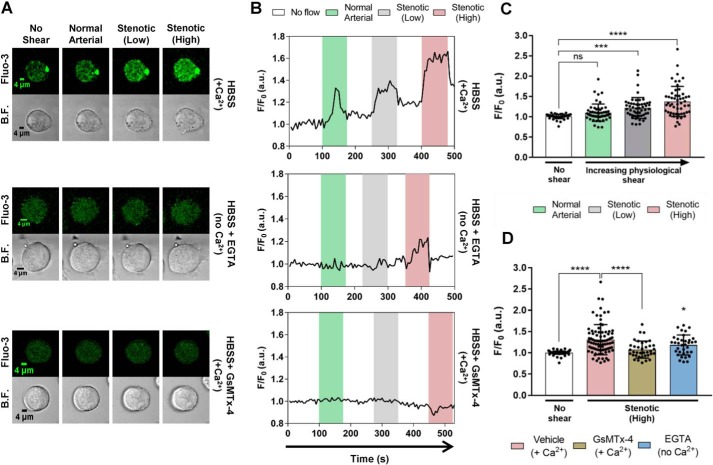
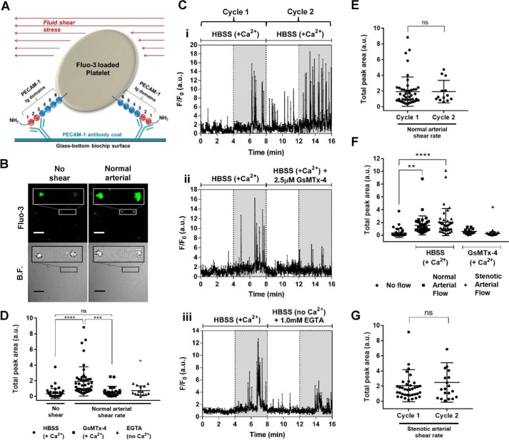
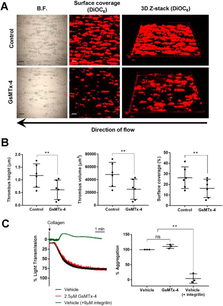
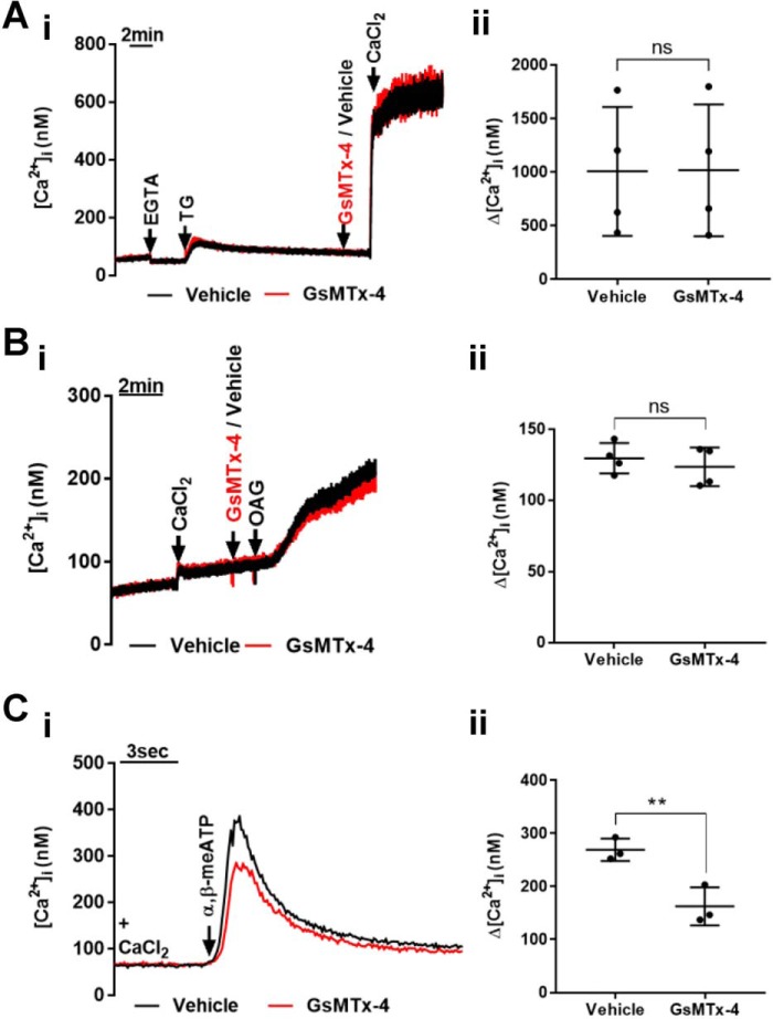
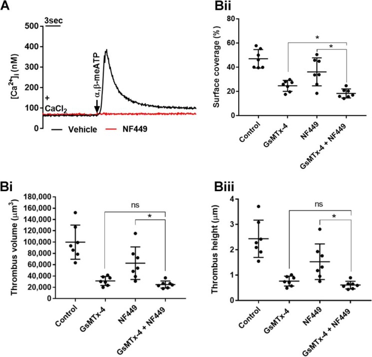
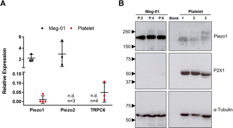
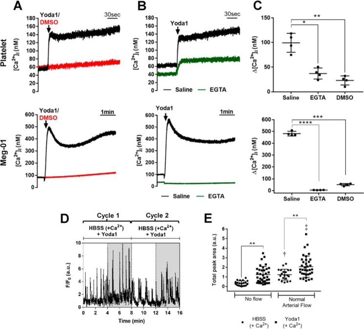
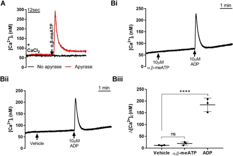
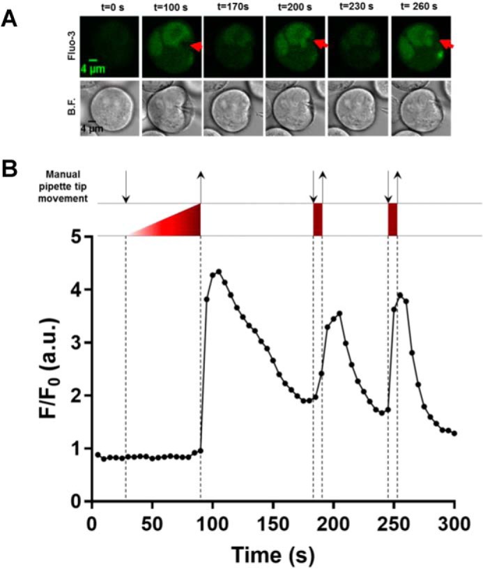
The role of mechanosensitive (MS) Ca2+-permeable ion channels in platelets is unclear, despite the importance of shear stress in platelet function and life-threatening thrombus formation. We therefore sought to investigate the expression and functional relevance of MS channels in human platelets. The effect of shear stress on Ca2+ entry in human platelets and Meg-01 megakaryocytic cells loaded with Fluo-3 was examined by confocal microscopy. Cells were attached to glass coverslips within flow chambers that allowed applications of physiological and pathological shear stress. Arterial shear (1002.6 s-1) induced a sustained increase in [Ca2+] i in Meg-01 cells and enhanced the frequency of repetitive Ca2+ transients by 80% in platelets. These Ca2+ increases were abrogated by the MS channel inhibitor Grammostola spatulata mechanotoxin 4 (GsMTx-4) or by chelation of extracellular Ca2+ Thrombus formation was studied on collagen-coated surfaces using DiOC6-stained platelets. In addition, [Ca2+] i and functional responses of washed platelet suspensions were studied with Fura-2 and light transmission aggregometry, respectively. Thrombus size was reduced 50% by GsMTx-4, independently of P2X1 receptors. In contrast, GsMTx-4 had no effect on collagen-induced aggregation or on Ca2+ influx via TRPC6 or Orai1 channels and caused only a minor inhibition of P2X1-dependent Ca2+ entry. The Piezo1 agonist, Yoda1, potentiated shear-dependent platelet Ca2+ transients by 170%. Piezo1 mRNA transcripts and protein were detected with quantitative RT-PCR and Western blotting, respectively, in both platelets and Meg-01 cells. We conclude that platelets and Meg-01 cells express the MS cation channel Piezo1, which may contribute to Ca2+ entry and thrombus formation under arterial shear.

Keywords: Piezo1; calcium; ion channel; mechanotransduction; platelet; shear stress.


Conflict of interest statement

The authors declare that they have no conflicts of interest with the contents of this article

Figures

Figure 1.
Figure 1.
Fluid shear stress-dependent Ca2+ influx in Meg-01 cells is inhibited by GsMTx-4 and chelation of extracellular Ca2+. A and B, representative images (A) and F/F0 fluorescence recordings (B) of single Meg-01 cells exposed to arterial and two levels of stenotic shear in HBSS with Ca2+, without Ca2+ (EGTA), and with GsMTx-4 in the presence of Ca2+. C, mean peak F/F0 increases (n = 53 cells) in response to different shear levels in the presence of extracellular Ca2+. D, mean peak F/F0 increases under no flow conditions (n = 113 cells) and at the high stenotic shear rate with (n = 85 cells) and without extracellular Ca2+ (n = 35 cells) and with GsMTx-4 in the presence of Ca2+ (n = 37 cells). ****, p < 0.0001; *, p < 0.05; **, p < 0.01. All cells were from cell culture passages 1–11. B.F., bright field.
Figure 2.
Figure 2.
Fluid shear stress induces Ca2+ transients in single platelets that are inhibited by GsMTx-4 and chelation of extracellular Ca2+. A, a cartoon representation of single platelet attachment to PECAM-1 antibody-coated biochip surface via the Ig domains 1 and 2 of the platelet PECAM-1. B, representative Fluo-3 fluorescence and bright field (B.F.) images of individual Fluo-3-loaded attached platelets before and during exposure to arterial shear. Scale bars, 20 μm. The magnified rectangular sections have been enlarged 3-fold. C, representative F/F0 Fluo-3 recordings in single platelets during no applied shear stress (white regions) and normal arterial shear (gray regions). Two successive cycles of 4 min without shear followed by 4 min of arterial shear were applied, in which the second cycle was used to compare the control conditions (i.e. HBSS + Ca2+ only) (panel i), with the effect of GsMTx-4 (panel ii), or removal of extracellular Ca2+ (panel iii). D–G, average Ca2+ increases, calculated as the 4-min F/F0 integral of all [Ca2+]i transients. D, responses in the absence of shear and during arterial shear, in the presence of extracellular Ca2+ with and without GsMTx-4 and in the absence of extracellular Ca2+ (n = 46, 46, 23, and 14 cells in no shear, HBSS, GsMTx-4, and EGTA, respectively). *, p < 0.05, compared with HBSS-only control under shear. E, comparison of Ca2+ responses during cycles 1 and 2 of normal arterial flow with Ca2+-containing HBSS only (n = 46 and 13 cells, respectively). No significant difference was found between F/F0 integrals of the calcium transients from cycles 1 and 2. F, Ca2+ responses in Ca2+-containing HBSS in the absence of shear (no applied flow) and during arterial stenotic shear with and without GsMTx-4 (n = 36, 36, 38, 13, and 13 cells in no applied flow, HBSS normal arterial, HBSS stenotic arterial, GsMTx-4 normal arterial, and GsMTx-4 stenotic arterial flow conditions, respectively). *, p < 0.05, compared with HBSS-only control under stenotic shear. G, no significant difference was found between F/F0 integrals of the calcium transients from cycles 1 and 2 of stenotic arterial flow, using Ca2+-containing HBSS only (n = 38 and 20 cells, respectively). ****, p < 0.0001; ***, p < 0.001; **, p < 0.01. ns, not significant.
Figure 3.
Figure 3.
Collagen-induced thrombus formation but not platelet aggregation is inhibited by GsMTx-4. A, representative images of surface coverage and 3D Z-stacks for thrombi formed by DiOC6-stained platelets on a collagen surface under control and GsMTx-4-pretreated conditions. Scale bars, 20 μm. B.F., bright field. B, average values (n = 6) for thrombus height, thrombus volume, and surface coverage under control and GsMTx-4-treated conditions. C, collagen-evoked aggregation under control and GsMTx-4-treated conditions. Integrilin treatment was performed as a control to demonstrate that aggregation is abolished by inhibition of the αIIbβ3 integrin. Representative light transmission traces are shown in the left panel, and average maximal light transmission responses expressed as percentages of aggregation are shown in the right panel (n = 3). **, p < 0.01. ns, not significant.
Figure 4.
Figure 4.
Effect of GsMTx-4 on Ca2+ entry via TRPC6, P2X1, and store-operated channels in platelets. A–C, representative [Ca2+]i recordings (left panels) and average peak [Ca2+]i responses (right panels) for store-operated (n = 4) (A), TRPC6 (n = 4) (B), and P2X1 cation channels (n = 3) (C) in suspensions of platelets in the presence and absence of GsMTx-4. Store-operated Ca2+ entry was assessed by addition of 1.26 mm CaCl2 15 min after treatment with the SERCA inhibitor thapsigargin. TRPC6 was activated using the diacylglycerol analogue OAG. P2X1 was activated with the non-hydrolyzable ATP analogue α,β-meATP (10 μm). **, p < 0.01; ns, not significant.
Figure 5.
Figure 5.
GsMTx-4 inhibits thrombus formation independently of P2X1 receptors. A, P2X1-dependent Ca2+ entry (α,β-meATP, 10 μm) in platelet suspensions is completely inhibited by 1 μm NF449. B, effect of 2.5 μm GsMTx-4 and 1 μm NF449, individually and combined, on thrombi formed on a collagen surface. The average values are shown (n = 7) for thrombus volume (panel i), surface coverage (panel ii), and thrombus height (panel iii). *, p < 0.05. ns, not significant.
Figure 6.
Figure 6.
Mechanosensitive ion channel expression in human platelets and the Meg-01 cell line. A, relative expression of mRNA transcripts for three MS cation channels (Piezo1, Piezo2, and TRPC6) in human platelets and the Meg-01 cell line, relative to GAPDH. n.d., not detected. The values are shown only for detectable levels of expression. B, Western blots for Piezo1 (233 kDa) and P2X1 receptors (55 kDa) in Meg-01 and human platelet lysates, compared with α-tubulin housekeeping control (60 kDa). The sizes (in kDa) and positions of protein standards are indicated with arrowheads. Meg-01 samples were from three different culture passages (lanes P:2, P:4, and P:6), and platelet samples were from three different donors (lanes 1, 2, and 3). The blank lane lacked protein lysate.
Figure 7.
Figure 7.
The Piezo1 agonist Yoda1 induced increases in [Ca2+]i in platelets and Meg-01 cells. A–C, [Ca2+]i responses to Yoda1 (25 μm) assessed in stirred Fura-2-loaded washed suspensions of platelets (top panels) and Meg-01 cells (bottom panels). A and B show representative recordings, and C shows the average peak [Ca2+]i increases (n = 4) for Yoda1 in the presence of extracellular Ca2+ compared with its vehicle control (DMSO) and following removal of external Ca2+ (EGTA). D, representative intracellular Ca2+ recording (Fluo-3 F/F0 fluorescence) from a single platelet attached to a PECAM-1-coated glass coverslip in the presence of Yoda1 and exposed to two cycles of no flow (white regions) and arterial shear (gray regions). See Fig. 2C, (panel i) for the control trace. E, average Ca2+ increases above baseline (F/F0·4 min) in the presence and absence of Yoda1 under conditions of no flow and normal arterial shear (n = 20, 35, 20, and 35 cells in HBSS no flow, Yoda1 no flow, HBSS normal arterial, and Yoda1 normal arterial conditions, respectively). **, p < 0.01; †, p < 0.01 compared with no flow, in the presence of HBSS; ‡, p < 0.001 compared with no flow in presence of Yoda1. Apyrase was omitted from the extracellular buffer to avoid P2X1 receptor responses (see Fig. 8).
Figure 8.
Figure 8.
Assessment of P2X1 activity in Fura-2-loaded platelet and Meg-01 cell suspensions in the presence and absence of extracellular apyrase. A, selective activation of platelet P2X1 channels using α,β-meATP in the presence and absence of 0.32 unit/ml apyrase. B, representative [Ca2+]i recordings (panels i and ii) and average Ca2+ increases (panel iii) demonstrating that α,β-meATP does not induce [Ca2+]i elevations in Meg-01 cells similar to vehicle control, indicating no P2X1 activity in these cells. As a positive control, the addition of a supramaximal concentration of ADP is shown to cause sharp [Ca2+]i elevations indicating intact P2Y responses. ****, p < 0.0001. ns, not significant.
Figure 9.
Figure 9.
Mechanical stimulation of Meg-01 cells with a glass pipette tip results in [Ca2+]i elevations. A, representative images of a Fluo-3-loaded Meg-01 cell at specified time points before and during [Ca2+]i elevations stimulated by depression of the plasma membrane with a blunt-ended glass micropipette. The extracellular saline (HBSS) contained 1.26 mm Ca2+. The red arrowheads indicate the positions and the directions in which the glass probe was applied. Similar responses were obtained from 14 Meg-01 cells from three different cultures. B.F., bright field. B, the F/F0 fluorescence recording of the Meg-01 cell shown in A. The downward arrows indicate when a push was applied onto the cell, and upward arrows indicate release of push. The regions enclosed with dashed lines represent the duration of a mechanical push by the glass probe.

References

    1. Schneider S. W., Nuschele S., Wixforth A., Gorzelanny C., Alexander-Katz A., Netz R. R., and Schneider M. F. (2007) Shear-induced unfolding triggers adhesion of von Willebrand factor fibers. Proc. Natl. Acad. Sci. U.S.A. 104, 7899–7903 - PMC - PubMed
    1. Li Z., Delaney M. K., O'Brien K. A., and Du X. (2010) Signaling during platelet adhesion and activation. Arterioscler. Thromb. Vasc. Biol. 30, 2341–2349 - PMC - PubMed
    1. Varga-Szabo D., Braun A., and Nieswandt B. (2009) Calcium signaling in platelets. J. Thromb. Haemost. 7, 1057–1066 - PubMed
    1. Mahaut-Smith M. P. (2012) The unique contribution of ion channels to platelet and megakaryocyte function. J. Thromb. Haemost. 10, 1722–1732 - PubMed
    1. Wright J. R., Amisten S., Goodall A. H., and Mahaut-Smith M. P. (2016) Transcriptomic analysis of the ion channelome of human platelets and megakaryocytic cell lines. Thromb. Haemost. 116, 272–284 - PMC - PubMed