. 2017 Jun 9;292(23):9830-9839.
doi: 10.1074/jbc.M116.763128. Epub 2017 Apr 17.

The deubiquitinating enzyme Usp14 allosterically inhibits multiple proteasomal activities and ubiquitin-independent proteolysis

Affiliations

The deubiquitinating enzyme Usp14 allosterically inhibits multiple proteasomal activities and ubiquitin-independent proteolysis

Hyoung Tae Kim et al. J Biol Chem. .

Abstract

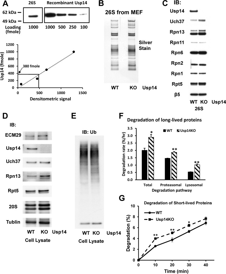
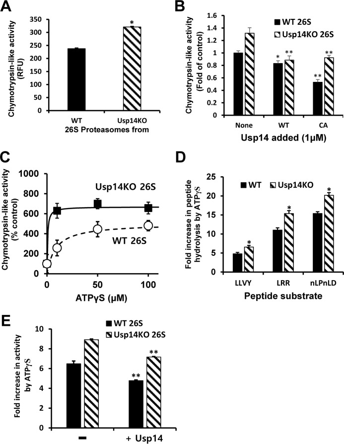
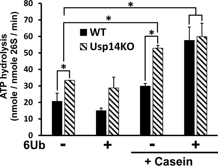
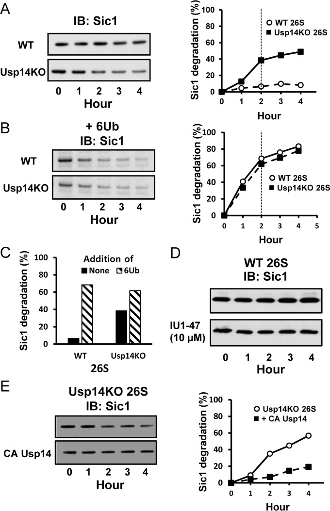
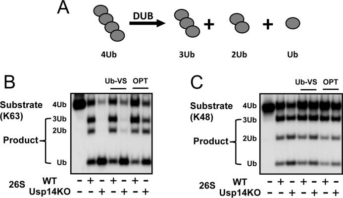
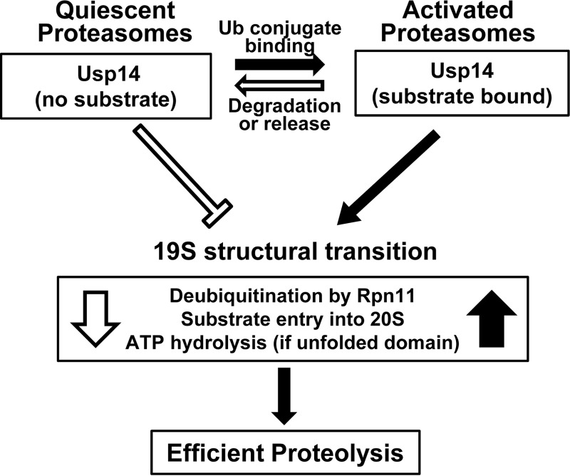
The proteasome-associated deubiquitinating enzyme Usp14/Ubp6 inhibits protein degradation by catalyzing substrate deubiquitination and by poorly understood allosteric actions. However, upon binding a ubiquitin chain, Usp14 enhances proteasomal degradation by stimulating ATP and peptide degradation. These studies were undertaken to clarify these seemingly opposite regulatory roles of Usp14 and their importance. To learn how the presence of Usp14 on 26S proteasomes influences its different activities, we compared enzymatic and regulatory properties of 26S proteasomes purified from wild-type mouse embryonic fibroblast cells and those lacking Usp14. The proteasomes lacking Usp14 had higher basal peptidase activity than WT 26S, and this activity was stimulated to a greater extent by adenosine 5'-O-(thiotriphosphate) (ATPγS) than with WT particles. These differences were clear even though Usp14 is present on only a minor fraction (30-40%) of the 26S in WT mouse embryonic fibroblast cells. Addition of purified Usp14 to the WT and Usp14-defficient proteasomes reduced both their basal peptidase activity and the stimulation by ATPγS. Usp14 inhibits these processes allosterically because a catalytically inactive Usp14 mutant also inhibited them. Proteasomes lacking Usp14 also exhibited greater deubiquitinating activity by Rpn11 and greater basal ATPase activity than WT particles. ATP hydrolysis by WT proteasomes is activated if they bind a ubiquitinated protein, which is loosely folded. Surprisingly, proteasomes lacking Usp14 could be activated by such proteins even without a ubiquitin chain present. Furthermore, proteasomes lacking Usp14 are much more active in degrading non-ubiquitinated proteins (e.g. Sic1) than WT particles. Thus, without a ubiquitinated substrate present, Usp14 suppresses multiple proteasomal activities, especially basal ATP consumption and degradation of non-ubiquitinated proteins. These allosteric effects thus reduce ATP hydrolysis by inactive proteasomes and nonspecific proteolysis and enhance proteasomal specificity for ubiquitinated proteins.

Keywords: ATPase; Usp14; allosteric regulation; deubiquitylation (deubiquitination); gate opening; peptidase; proteasome; protein degradation.


Conflict of interest statement

The authors declare that they have no conflicts of interest with the contents of this article

Figures

Figure 1.
Figure 1.
MEF cells lacking Usp14 have increased content of Rpn13 and Uch37 in the 26S proteasomes. Proteasomes were purified from WT and Usp14KO MEFs using the UBL method (see “Experimental procedures”). Concentrations of each proteasome preparation were determined using the BCA method, and molarities were calculated using 2.5 MDa as the molecular mass of 26S proteasomes. A, Usp14 is a substoichiometric component of WT MEF 26S. The content of Usp14 in 1 pmol of WT 26S proteasomes was determined by Western blotting using recombinant Usp14 as the standard. The content of Usp14 in 1 pmol of WT MEF 26S was calculated as 380 fmol. Upper panel, Western blot. Lower panel, quantitation from upper panel using ImageJ software. B, SDS-PAGE of purified 26S proteasomes from WT and Usp14KO MEFs. Proteins were silver-stained after electrophoresis. C, the contents of Uch37 and Rpn13 were increased in the purified 26S proteasomes from Usp14KO MEFs even though Usp14KO 26S contains the same levels of other subunits as WT 26S. Levels of proteins in 26S proteasomes purified from WT and Usp14KO MEF were determined by Western blotting after SDS-PAGE. D, the expression of Rpn13 was increased in Usp14KO MEFs. Levels of proteasome subunits were determined by Western blotting of cell lysates of WT and Usp14KO MEFs after SDS-PAGE. Unlike the purified proteasomes, only the content of Rpn13, but not that of Uch37, was increased in Usp14KO MEF cells. Therefore, this increase in Uch37 content must be due to increased binding of Rpn13. The content of tubulin was monitored as an input control. E, the cellular content of Ub conjugates did not change significantly in Usp14KO MEFs. The levels of Ub conjugates were determined in lysates of WT and Usp14KO MEFs by Western blotting after SDS-PAGE. F, MEF cells lacking Usp14 degrade long-lived cell proteins faster than WT MEFs. WT and Usp14KO MEF cells were labeled with [3H]phenylalanine (5 μCi/ml) for 24 h. After washing the cells with PBS and then chase medium (DMEM containing 2 mg/ml nonradioactive phenylalanine and 100 μm cycloheximide), cells were grown for a further 2 h to let short-lived labeled proteins be degraded. Then cells were grown in the chase medium with either DMSO (control) or 10 μm bortezomib/Velcade dissolved in DMSO to block proteasome activity. After 1 h of inhibitor treatment, degradation of cellular proteins was measured in the chase medium for up to 4 h. Proteasomal degradation rates were calculated by subtracting the degradation rate with bortezomib treatment from the total degradation rate. Error bars represent S.D. The remaining proteasome-independent proteolysis represents lysosomal proteolysis. *, p < 0.05; **, p < 0.01 compared with WT MEF by Student's t test. G, MEF cells lacking Usp14 degrade short-lived cell proteins faster than WT MEFs. WT and Usp14KO MEF cells were labeled with [3H]phenylalanine (10 μCi/ml) for 20 min. After washing the cells as in F, cells were incubated for 10 min in the chase medium. Degradation of cellular proteins was then measured in the chase medium for up to 40 min. Error bars represent S.D. *, p < 0.05; **, p < 0.01 compared with WT MEF by Student's t test. n = 6. IB, immunoblotting.
Figure 2.
Figure 2.
Usp14 reduces peptidase activities in the proteasome and the activation of peptide entry by ATPγS. Peptidase activities of purified WT or Usp14KO 26S were assayed by measuring hydrolysis of fluorogenic peptide-amc substrates (10 μm; Suc-LLVY-amc for chymotrypsin-like activity, Boc-LRR-amc for trypsin-like activity, and Ac-nLPnLD-amc for caspase-like activity) in the presence of 100 μm ATP or ATPγS. The basal peptidase activity (control) of the proteasomes was measured in the presence of ATP. Error bars represent S.D. of at least three independent measurements. A, chymotrypsin-like activity of 26S purified from Usp14KO cells (4 nm) is consistently greater (35%) than that of WT 26S in the presence of ATP (100 μm) even though only a small fraction (∼40%) of the proteasomes from WT cells contained Usp14 (Fig. 1A). Cleavage of the fluorogenic substrate Suc-LLVY-amc (10 μm) by each type of proteasomes was monitored. *, p < 0.05 compared with WT 26S by Student's t test. B, binding of full-length recombinant Usp14 protein to the 26S particles inhibits the chymotrypsin-like activity of WT and Usp14KO 26S. Purified proteasomes (100 nm) were preincubated for 10 min at room temperature with bacterially expressed full-length WT or an inactive Cys → Ala (CA) mutant Usp14 (1 μm) before the start of the assays. *, p < 0.05; **, p < 0.01 compared with control proteasomes (without Usp14 added) by Student's t test. C, increasing amounts of ATPγS stimulate the chymotrypsin-like activity of both WT and Usp14KO proteasomes in a saturable manner. Both the maximal stimulation by ATPγS and the apparent affinity for ATPγS were greater for Usp14KO 26S than for the WT particles. D, ATPγS (100 μm) markedly stimulated all three peptidase activities of both WT and Usp14KO proteasomes (4 nm) but consistently caused a 30–40% larger stimulation of these activities in the Usp14KO 26S. *, p < 0.05 compared with WT 26S by Student's t test. E, addition of Usp14 reduced the stimulation of the chymotrypsin-like activity by ATPγS. After incubation of purified proteasomes (100 nm) with a molar excess of Usp14 (1 μm) at room temperature for 10 min, the chymotrypsin-like activities of WT and Usp14KO 26S with ATPγS (100 μm) were assayed as in D. **, p < 0.01 compared with control samples without Usp14 added by Student's t test. RFU, relative fluorescence units.
Figure 3.
Figure 3.
ATPases of Usp14KO proteasomes, unlike those of WT 26S, are stimulated by unfolded proteins in the absence of a Ub chain. ATP hydrolysis by 26S proteasomes (20 nm) was measured using the malachite green assay (54). The basal ATPase activity of Usp14KO 26S was consistently (60%) higher than that of WT 26S. Upon addition of casein (1 μm) and a hexa-Ub chain (1 μm), which together mimic the binding of a Ub conjugate (22), ATP hydrolysis of WT 26S increased 2–3-fold and resembled the ATPase activity of Usp14KO 26S under the same conditions. However, hexa-Ub alone did not increase ATP hydrolysis of either type of proteasomes. ATP hydrolysis by Usp14KO 26S, but not that by WT 26S, was enhanced by addition of casein alone and reached the same level as seen with 26S upon binding both a Ub chain and casein. 6Ub, linear hexa-Ub chain. *, p < 0.05 compared with control by Student's t test. Error bars represent S.D.
Figure 4.
Figure 4.
Usp14 reduces proteasomal degradation of unstructured proteins lacking ubiquitination. Degradation of the intrinsically unstructured protein PY-Sic1 (100 nm) by 26S proteasomes (2 nm) was assayed by Western blotting. A and B, left panels, after reaction for the indicated times, the remaining Sic1 was analyzed by Western blotting. Right panels, degradation of Sic1 was plotted by measuring Sic1 density using ImageJ software. Each point is representative of at least two independent experiments. A, unlike 26S proteasomes from WT cells, Usp14KO 26S proteasomes degrade non-ubiquitinated PY-Sic1. These data are representative of three independent experiments. B, although the basal degradation of PY-Sic1 by 26S from Usp14KO MEFs was much higher than that of WT proteasomes, the degradation rates by both types of 26S particles were quite similar in the presence of linear hexa-Ub chain (1 μm). Thus, the Ub chains stimulate degradation by the WT proteasomes much more than by the Usp14KO 26S. Similar data were obtained in three independent experiments. 26S proteasomes were incubated with the Ub chain at room temperature for 15 min before the reaction started. C, the dependence of proteasomal degradation of PY-Sic1 on the presence of a Ub chain was much greater in WT than Usp14KO 26S. Addition of Ub chain stimulated degradation 10-fold in WT 26S and 60% in Usp14KO particles. Degradation rates were compared after incubation for 2 h when reaction rates slowed below initial rates (at the vertical line in A and B). D, although knock-out of Usp14 stimulates the degradation of PY-Sic1, a Usp14-specific inhibitor IU1 (10 μm) does not stimulate the degradation of PY-Sic1 by the purified WT 26S particles nor does it affect peptide hydrolysis (data not shown) even though knock-out of Usp14 stimulates their degradation. E, addition of inactive Cys → Ala (CA) Usp14 mutant inhibits degradation of Sic1 by Usp14KO 26S. These proteasomes were preincubated with Usp14 as in Fig. 2B before the degradation reactions. These data are representative of two independent experiments. IB, immunoblotting.
Figure 5.
Figure 5.
Usp14 inhibits deubiquitination by Rpn11. A, disassembly of tetra-Ub chain (368 nm) by WT or Usp14KO 26S (5 nm) was assayed after incubation at 37 °C for 20 min. The generation of tri-, di-, and mono-Ubs was analyzed by Western blotting after electrophoresis. B and C, Usp14KO MEF 26S proteasomes disassembled tetra-Ub chains much faster than WT 26S particles. Although Ub-VS (1.5 μm) did not inhibit Ub chain hydrolysis, o-phenanthroline (OPT) (1 mm) markedly inhibits Ub chain hydrolysis and reduces deubiquitination by Usp14KO 26S to the level of WT 26S. Thus, this process involves the Zn2+ metalloprotease Rpn11. Similar results were also obtained with Lys-63 tetra-Ub chains (B) and Lys-48 tetra-Ub chains (C) as substrates and were obtained in at least three independent experiments.
Figure 6.
Figure 6.
Summary of Usp14's allosteric regulation of proteasomal degradation. Without a Ub conjugate bound, proteasomes are relatively inactive, and Usp14 inhibits ATP hydrolysis, substrate entry into the 20S, and deubiquitination by Rpn11. These actions reduce futile ATP consumption and the degradation of non-ubiquitinated proteins, which occur in the Usp14KO 26S but not in WT 26S. However, upon Ub chain binding, Usp14 (and the 19S particle) undergoes a major structural transition and thus activates Rpn11, peptide entry into 20S, and ATP hydrolysis (if an unfolded protein also is bound). These actions promote efficient degradation of ubiquitinated substrates. This activated state should be maintained as long as Usp14 binds a Ub chain. These processes return to the quiescent basal state when the substrate is degraded or is deubiquitinated and dissociates. These allosteric actions are not dependent on Usp14's catalytic activity, which functions to degrade the Ub chain and thus limits the duration of the active state and the substrate's “dwell time” on the 19S complex.

Similar articles

Cited by

References

    1. Finley D. (2009) Recognition and processing of ubiquitin-protein conjugates by the proteasome. Annu. Rev. Biochem. 78, 477–513 - PMC - PubMed
    1. Smith D. M., Benaroudj N., and Goldberg A. (2006) Proteasomes and their associated ATPases: a destructive combination. J. Struct. Biol. 156, 72–83 - PubMed
    1. Kisselev A. F., Garcia-Calvo M., Overkleeft H. S., Peterson E., Pennington M. W., Ploegh H. L., Thornberry N. A., and Goldberg A. L. (2003) The caspase-like sites of proteasomes, their substrate specificity, new inhibitors and substrates, and allosteric interactions with the trypsin-like sites. J. Biol. Chem. 278, 35869–35877 - PubMed
    1. Groll M., and Huber R. (2003) Substrate access and processing by the 20S proteasome core particle. Int. J. Biochem. Cell Biol. 35, 606–616 - PubMed
    1. Groll M., Bajorek M., Köhler A., Moroder L., Rubin D. M., Huber R., Glickman M. H., and Finley D. (2000) A gated channel into the proteasome core particle. Nat. Struct. Biol. 7, 1062–1067 - PubMed

MeSH terms

LinkOut - more resources