. 2017 May 2;114(18):E3642-E3651.
doi: 10.1073/pnas.1619266114. Epub 2017 Apr 17.

Drebrin restricts rotavirus entry by inhibiting dynamin-mediated endocytosis

Affiliations

Drebrin restricts rotavirus entry by inhibiting dynamin-mediated endocytosis

Bin Li et al. Proc Natl Acad Sci U S A. .

Abstract

Despite the wide administration of several effective vaccines, rotavirus (RV) remains the single most important etiological agent of severe diarrhea in infants and young children worldwide, with an annual mortality of over 200,000 people. RV attachment and internalization into target cells is mediated by its outer capsid protein VP4. To better understand the molecular details of RV entry, we performed tandem affinity purification coupled with high-resolution mass spectrometry to map the host proteins that interact with VP4. We identified an actin-binding protein, drebrin (DBN1), that coprecipitates and colocalizes with VP4 during RV infection. Importantly, blocking DBN1 function by siRNA silencing, CRISPR knockout (KO), or chemical inhibition significantly increased host cell susceptibility to RV infection. Dbn1 KO mice exhibited higher incidence of diarrhea and more viral antigen shedding in their stool samples compared with the wild-type littermates. In addition, we found that uptake of other dynamin-dependent cargos, including transferrin, cholera toxin, and multiple viruses, was also enhanced in DBN1-deficient cells. Inhibition of cortactin or dynamin-2 abrogated the increased virus entry observed in DBN1-deficient cells, suggesting that DBN1 suppresses dynamin-mediated endocytosis via interaction with cortactin. Our study unveiled an unexpected role of DBN1 in restricting the entry of RV and other viruses into host cells and more broadly to function as a crucial negative regulator of diverse dynamin-dependent endocytic pathways.

Keywords: drebrin; endocytosis; rotavirus.


Conflict of interest statement

The authors declare no conflict of interest.

Figures

Fig. 1.
Fig. 1.
VP4 proteomic network reveals cytoskeleton-binding proteins that modulate RV infection. (A) Interactome of the bait viral protein (VP4) and high-confidence host binding proteins. Solid lines with arrowheads represent interactions identified in this study. The width of lines corresponds to the strength of interaction detected in the IP–MS experiment. Dotted lines represent publicly curated protein–protein interactions. The proteins studied in this paper are highlighted by yellow nodes. (B) Pie chart of PANTHER functional classification of VP4-interacting proteins shown in A. (C) HEK293 cells were transfected with indicated siRNA for 48 h and infected with simian RV RRV strain at MOI = 1 for 24 h. Levels of RV replication were measured by RT-qPCR examining the expression of viral gene VP7, normalized to that of GAPDH. The genes studied in this paper are outlined by red boxes. (D) Same experiment as in C except that viral gene NSP5 was measured instead of VP7 by RT-qPCR and virus titer in the supernatants was determined by a focus-forming unit (FFU) assay. For C and D, experiments were repeated at least five times. Data are represented as mean ± SEM. Statistical significance is determined by Student’s t test (**P ≤ 0.01; ***P ≤ 0.001).
Fig. 2.
Fig. 2.
Drebrin deficiency results in enhanced RV infection. (A) Lysates of HEK293 cells stably expressing a GFP-tagged RRV VP4 were stimulated with or without doxycycline (Dox, 1 μg/mL) for 24 h, then subjected to IP using α-GFP antibody, and analyzed with Western blot using indicated antibodies. Bottom panels are 10% input. (B) HEK293 cells were infected with RRV at MOI = 3 for 24 h and analyzed by confocal microscopy for the localization of VP4 (green), DBN1 (red), actin (phalloidin, white), and nucleus (DAPI, blue). Colocalization (yellow) is highlighted by white arrowheads. Panels are single z slices. (Scale bar, 8 μm.) (C) WT and two individual clones of DBN1 knockout (KO) HEK293 cells were infected with RRV at MOI = 1 for 24 h. Viral gene NSP5 expression was measured by RT-qPCR and normalized to that of GAPDH (Left). Virus particles in the supernatants were titrated by an FFU assay (Right). (D) DBN1 KO HEK293 cells were transfected with pCMV6-entry empty vector (ev), and Flag-tagged full-length DBN-A or DBN-E, and infected with RRV at MOI = 1 for 24 h. Viral gene NSP5 level was measured by RT-qPCR and normalized to that of GAPDH (Left) and lysates were harvested for Western blot using indicated antibodies (Right). (E) WT and DBN1 KO HEK293 cells were transfected with RV double-layered particles (DLPs) for 16 h and NSP5 expression was measured by RT-qPCR. (F) WT and DBN1 KO HEK293 cells were infected with RRV (MOI = 50) and then incubated at either 4 °C for 1 h or incubated at 4 °C for 1 h and then shifted to 37 °C for an additional 1 h. The level of viral gene NSP5 was measured by RT-qPCR and normalized to that of GAPDH, as an indicator of input viral genomes. For all figures, experiments were repeated at least three times. Data are represented as mean ± SEM. Statistical significance is determined by Student’s t test (**P ≤ 0.01; ***P ≤ 0.001; n.s., not significant).
Fig. 3.
Fig. 3.
Loss of drebrin leads to enhanced endocytosis of dynamin-dependent cargo. (A) WT and DBN1 KO HEK293 cells were incubated with FITC-conjugated transferrin (Tfn, 20 μg/mL) and fixed at indicated time points for confocal microscopy to examine intracellular transferrin (green) and nucleus (DAPI, blue). (Scale bar in panels and single z slices, 20 μm.) (B) WT and DBN1 KO HEK293 cells were incubated with FITC-conjugated cholera toxin subunit B (CTxB, 10 μg/mL) and fixed at indicated time points for confocal microscopy for internalized toxin (green) and nucleus (DAPI, blue). (Scale bar in panels and single z slices, 20 μm.) (C) WT and DBN1 KO HEK293 cells were infected with human adenovirus serotype 5 (HAdV5) or simian vacuolating virus 40 (SV40) at MOI = 0.1 for 1 h. Expression levels of hexon (HAdV5) and large T antigen (SV40) were measured by RT-qPCR and normalized to that of GAPDH. For all figures, experiments were repeated at least three times. Data are represented as mean ± SEM. Statistical significance is determined by Student’s t test (***P ≤ 0.001).
Fig. 4.
Fig. 4.
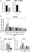
Drebrin inhibits dynamin-2–mediated endocytosis via interaction with cortactin. (A) WT and DBN1 KO HEK293 cells were treated with either vehicle control (DMSO) or dynasore (100 μM) for 30 min before RRV infection (MOI = 1). Total RNA was harvested at 1 hpi and NSP5 level was measured by RT-qPCR and normalized to that of GAPDH. (B) WT HEK293 cells were transfected with indicated siRNA for 48 h before infection with RRV at MOI = 1 for 1 h. Viral NSP5 level was measured by RT-qPCR and normalized to that of GAPDH. Dotted line denotes NSP5 level of cells transfected with ctrl siRNA (set at 0.984); cortactin (encoded by CTTN) is outlined by a red box. (C) DBN1 KO HEK293 cells were transfected with indicated siRNA (gelsolin, encoded by GSN) for 48 h and infected with RRV at MOI = 1 for 1 h. The level of viral NSP5 was measured by RT-qPCR and normalized to that of GAPDH. (D) HEK293 cells were transfected with the first siRNA for 48 h and then transfected with second siRNA for an additional 48 h before RRV infection (MOI = 1) for 1 h. The input viral genome was measured by NSP5 levels using RT-qPCR and normalized to that of GAPDH. For all figures, experiments were repeated at least three times. Data are represented as mean ± SEM. Statistical significance is determined by Student’s t test (*P ≤ 0.05; **P ≤ 0.01; ***P ≤ 0.001; n.s., not significant).
Fig. 5.
Fig. 5.
N-terminal domain of drebrin localizes to the actin cytoskeleton and coprecipitates with cortactin. (A) DBN1 KO HEK293 cells stably expressing indicated GFP-tagged DBN1 constructs or control EGFP were analyzed by confocal microscopy for the localization of DBN1 (green), actin (red), and nucleus (DAPI, blue). Colocalization (yellow) is highlighted by white arrowheads. (Full-length: DBN isoforms A and E; N terminus: amino acid 1–366; middle region: 233–317; C terminus: 319–707). (Scale bar in panels and single z slices, 40 μm.) (B) DBN1 KO HEK293 cells stably expressing indicated GFP-tagged DBN1 constructs or control EGFP were subject to IP using α-GFP antibody and analyzed by Western blot using indicated antibodies. The IP band intensities were normalized to endogenous CTTN levels in IP input and compared with that of DBN-A (lane 1), which was set as 1.00. Bottom panels are 10% input. (C) Reconstituted DBN1 KO HEK293 cells were infected with RRV at MOI = 1 for 24 h and examined by RT-qPCR for viral NSP5 expression, normalized to that of GAPDH. For all figures, experiments were repeated at least three times. Data are represented as mean ± SEM. Statistical significance is determined by Student’s t test (***P ≤ 0.001).
Fig. 6.
Fig. 6.
Drebrin knockout results in increased RV infection in vivo and in human enteroids. (A) Five-day-old C57BL/6 pups of indicated Dbn1 genotypes (WT, n = 3; Het, n = 7; KO, n = 3) were orally inoculated with 106 pfu of the simian RV RRV strain and monitored for the incidence of diarrhea for 7 d. (B) Quantification of A for the indicated days postinfection based on diarrheal severity parameters. (C) Fecal specimens were collected on the indicated days postinfection and subject to a standard plaque forming unit (pfu) assay to determine infectious virus particles per gram of stool samples. (D) Human intestinal organoids were treated with either vehicle control (DMSO) or BTP-2 at indicated concentrations for 30 min before infection with the human RV Wa strain (MOI = 1). Total RNA was harvested at 1 hpi and NSP5 level was measured by RT-qPCR and normalized to that of GAPDH. For D, experiments were repeated at least three times. Data are represented as mean ± SEM. Statistical significance is determined by Student’s t test (*P ≤ 0.05; **P ≤ 0.01; n.s., not significant).

Similar articles

Cited by

References

    1. Marsh M, editor. 2001. Endocytosis, Frontiers in Molecular Biology (Oxford University Press, Oxford)
    1. Ferguson SM, De Camilli P. Dynamin, a membrane-remodelling GTPase. Nat Rev Mol Cell Biol. 2012;13:75–88. - PMC - PubMed
    1. McMahon HT, Boucrot E. Molecular mechanism and physiological functions of clathrin-mediated endocytosis. Nat Rev Mol Cell Biol. 2011;12:517–533. - PubMed
    1. Cossart P, Helenius A. Endocytosis of viruses and bacteria. Cold Spring Harb Perspect Biol. 2014;6:6. - PMC - PubMed
    1. Mercer J, Schelhaas M, Helenius A. Virus entry by endocytosis. Annu Rev Biochem. 2010;79:803–833. - PubMed

Publication types