. 2017 May 2;114(18):4787-4792.
doi: 10.1073/pnas.1620289114. Epub 2017 Apr 17.

Sensorimotor-independent development of hands and tools selectivity in the visual cortex

Affiliations

Sensorimotor-independent development of hands and tools selectivity in the visual cortex

Ella Striem-Amit et al. Proc Natl Acad Sci U S A. .

Abstract

The visual occipito-temporal cortex is composed of several distinct regions specialized in the identification of different object kinds such as tools and bodies. Its organization appears to reflect not only the visual characteristics of the inputs but also the behavior that can be achieved with them. For example, there are spatially overlapping responses for viewing hands and tools, which is likely due to their common role in object-directed actions. How dependent is occipito-temporal cortex organization on object manipulation and motor experience? To investigate this question, we studied five individuals born without hands (individuals with upper limb dysplasia), who use tools with their feet. Using fMRI, we found the typical selective hand-tool overlap (HTO) not only in typically developed control participants but also in four of the five dysplasics. Functional connectivity of the HTO in the dysplasics also showed a largely similar pattern as in the controls. The preservation of functional organization in the dysplasics suggests that occipito-temporal cortex specialization is driven largely by inherited connectivity constraints that do not require sensorimotor experience. These findings complement discoveries of intact functional organization of the occipito-temporal cortex in people born blind, supporting an organization largely independent of any one specific sensory or motor experience.

Keywords: body image; brain development; motor deprivation; tool use; visual cortex.


Conflict of interest statement

The authors declare no conflict of interest.

Figures

Fig. 1.
Fig. 1.
Hand–tool visual cortex overlap does not require hands. (A) The hand and tool selectivity overlap (HTO) in the lateral occipito-temporal cortex is replicated in typically developed control subjects (n = 10, RFX GLM, P < 0.05, corrected for multiple comparisons). Yellow marks hand selectivity over feet, and purple marks tool selectivity over nonmanipulable artifacts. (B) The HTO, in the occipito-temporal cortex, can be found at the individual level in the majority (7 of 10) of the controls (for cortical surface views, see Fig. S2). (C) A majority of the dysplasics (4 of 5) born without hands show an overlap between hand and tool selectivity in the visual cortex. This suggests that motor experience is not critical for the formation and specialization of the visual-cortex hand action-related representations. (D) The location (Left; spheres denote the location of individual subjects HTO peaks) and size (Right) of the HTO in the dysplasics (marked yellow) did not differ from that of the control subjects (marked blue). (E) In addition to the overlap between hand and tool selectivity, 2 of the dysplasics (2 of 5) and 1 of the controls (1 of 10) show an overlap of feet and tools selectivity [a foot–tool overlap (FTO)]. This potentially suggests visual cortex organization is also somewhat plastic to changes due to different individual sensorimotor experience.
Fig. S1.
Fig. S1.
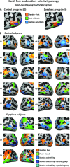
Hand, feet, and motion selectivity occupy distinct cortical regions. (A) Hand (orange), foot (green), and motion selectivity (blue) are viewed on the lateral occipito-temporal cortex in both groups. Hand and foot selectivity are compared on a winner-takes-all approach (each condition is compared with baseline at P < 0.05, FDR corrected, and the preferred body part is then selected for each vertex), revealing an inferior and posterior (dorsal) area, which show a preference for viewing feet over viewing hands in both groups. Motion selectivity was measured with an additional localizer (Materials and Methods; P < 0.05, FDR corrected). Importantly, hands- and feet-preferential responses do not all result from an overlap with motion-selective regions. (B) Hand (orange; hands > feet), foot (green; feet > hands), and motion (blue; moving > stationary rings) selectivity (measured by direct contrast) are depicted in individual subjects, largely reproducing the pattern seen in the group results of having distinct hand and foot preferences, which do not overlap with motion selectivity. For two dysplasic subjects who do not have individual motion selectivity localizers (D2, D3), group motion selectivity data from both groups is presented (in blue and cyan for the control and dysplasic groups, respectively; greatly overlapping with one another).
Fig. S2.
Fig. S2.
The hand–tool overlap (HTO) does not stem from motion selectivity. HTO (orange; hands > feet AND tools > nonmanipulable artifacts), foot–tool overlap (FTO) (green; feet > hands AND tools > nonmanipulable artifacts), and motion selectivity (blue; moving > stationary rings) are viewed on the lateral occipito-temporal cortex in each of the individual subjects (P < 0.05, corrected). For two dysplasic subjects who do not have individual motion selectivity localizers (D2, D3), group motion selectivity data from both groups is presented (in blue and cyan for the control and dysplasic groups, respectively; greatly overlapping with one another). The HTOs and FTOs do not overlap with motion selectivity. Therefore, general motion selectivity cannot account for the hand and tool conjoint selectivity in the dysplasics.
Fig. 2.
Fig. 2.
Connectivity from the occipito-temporal cortex reflects intact visual mechanisms alongside their plasticity. (A) Functional connectivity from the HTO in the control subjects (probabilistic mapping of the functional connectivity of individual subjects, reflecting the percentage of subjects showing this pattern) replicates the network of visual cortical regions engaged in visuomotor tool use processing, extending to the sensorimotor cortex of the hand region (independent localizer marked in white). (B) A highly similar network is connected to the HTO of the dysplasics, across the ventral and dorsal visual streams. One difference between the groups can be seen in the absence of functional connectivity to the primary sensorimotor cortex in the dysplasics. (C) A direct comparison of functional connectivity from the HTO between the dysplasics and control subjects was computed by Bayesian standardized difference test comparison of each dysplasic subject to the control group. The figure denotes a probabilistic mapping of differences, such that only two dysplasic subjects would have to show a difference from the control subjects for a voxel to be marked. Even at this permissive overlap threshold, relatively sparse small foci of differential functional connectivity can be found. These show heightened functional connectivity in the dysplasics in the visual cortex (including the superior parietal lobe), and decreased functional connectivity in the sensorimotor cortices, including the hand sensorimotor cortex (independent localizer: marked in white). (D) Functional connectivity was computed in three subjects, two dysplasics (D1 and D3), and one control (C9), which showed both an HTO and a foot–tool overlap (FTO). The dysplasics show functional connectivity from the FTO but little from HTO to their sensorimotor cortex [including their foot-selective regions, marked in white; these include atypical lateral foot responses (63); for full motor response maps, see Figs. S3 and S4]. In contrast, the control subject shows the reverse trend, of functional connectivity to the sensorimotor cortex only from her HTO. Therefore, the unique experience of the dysplasics in foot–tool use can manifest in functional connectivity linking the FTO to the motor cortex.
Fig. S3.
Fig. S3.
Motor responses in single subjects presented in Fig. 2. Motor foot selectivity (flexing the feet > flexing shoulders, stomach and mouth muscles; P < 0.05, corrected; depicted in orange) is presented for dysplasics D1 and D3, and control C9. The responses near the central sulcus are outlined and were used in Fig. 2 to identify the individual foot-selective regions. For subject C9, hand selectivity (flexing the hands > flexing shoulders, stomach and mouth muscles; P < 0.05, corrected; depicted in blue) is also presented.
Fig. S4.
Fig. S4.
The HTO responds to unseen hand movements in typically developed subjects, but not to foot movements. (A) Motor responses for hand movements in the control group (RFX, P < 0.05, corrected) are presented for two motor control experiments (vs. baseline; see Materials and Methods for detail). The left panel depicts activation during grasping and reaching actions, and the right panel depicts activation during performance of simple hand-flexing movements. Both movement patterns of the hands activate the HTO of the control subjects. (B) Foot actions in the control group (RFX, P < 0.05, corrected) do not activate the HTO, showing this region’s motor responsivity is specific and aligned to its visual selectivity. (C) Foot actions in the dysplasic group (P < 0.05, corrected) do not activate the HTO, suggesting the HTO’s development in the dysplasics does not result from foot-related motor imagery or simulation. Interestingly, the motor foot responses of the dysplasics spread to the lateral sensorimotor cortex, to areas usually occupied by hand motor selectivity, which are not activated by the control subjects (compare B). This is in accord with a past report of the overtake of the hand motor area in congenitally dysplasic individuals (63).

Similar articles

Cited by

References

    1. Kanwisher N. Functional specificity in the human brain: A window into the functional architecture of the mind. Proc Natl Acad Sci USA. 2010;107:11163–11170. - PMC - PubMed
    1. Caramazza A, Shelton JR. Domain-specific knowledge systems in the brain: The animate-inanimate distinction. J Cogn Neurosci. 1998;10:1–34. - PubMed
    1. Blundo C, Ricci M, Miller L. Category-specific knowledge deficit for animals in a patient with herpes simplex encephalitis. Cogn Neuropsychol. 2006;23:1248–1268. - PubMed
    1. Mahon BZ, Caramazza A. Concepts and categories: A cognitive neuropsychological perspective. Annu Rev Psychol. 2009;60:27–51. - PMC - PubMed
    1. Proklova D, Kaiser D, Peelen MV. Disentangling representations of object shape and object category in human visual cortex: The animate-inanimate distinction. J Cogn Neurosci. 2016;28:680–692. - PubMed

Publication types

LinkOut - more resources