. 2017 Apr 18:8:15054.
doi: 10.1038/ncomms15054.

A new inhibitor of the β-arrestin/AP2 endocytic complex reveals interplay between GPCR internalization and signalling

Affiliations

A new inhibitor of the β-arrestin/AP2 endocytic complex reveals interplay between GPCR internalization and signalling

Alexandre Beautrait et al. Nat Commun. .

Abstract

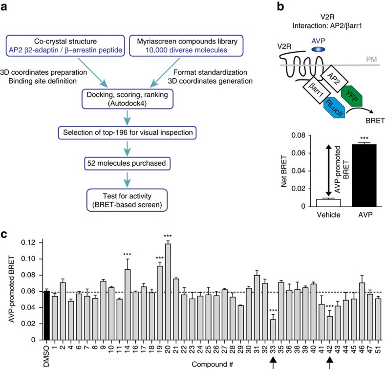
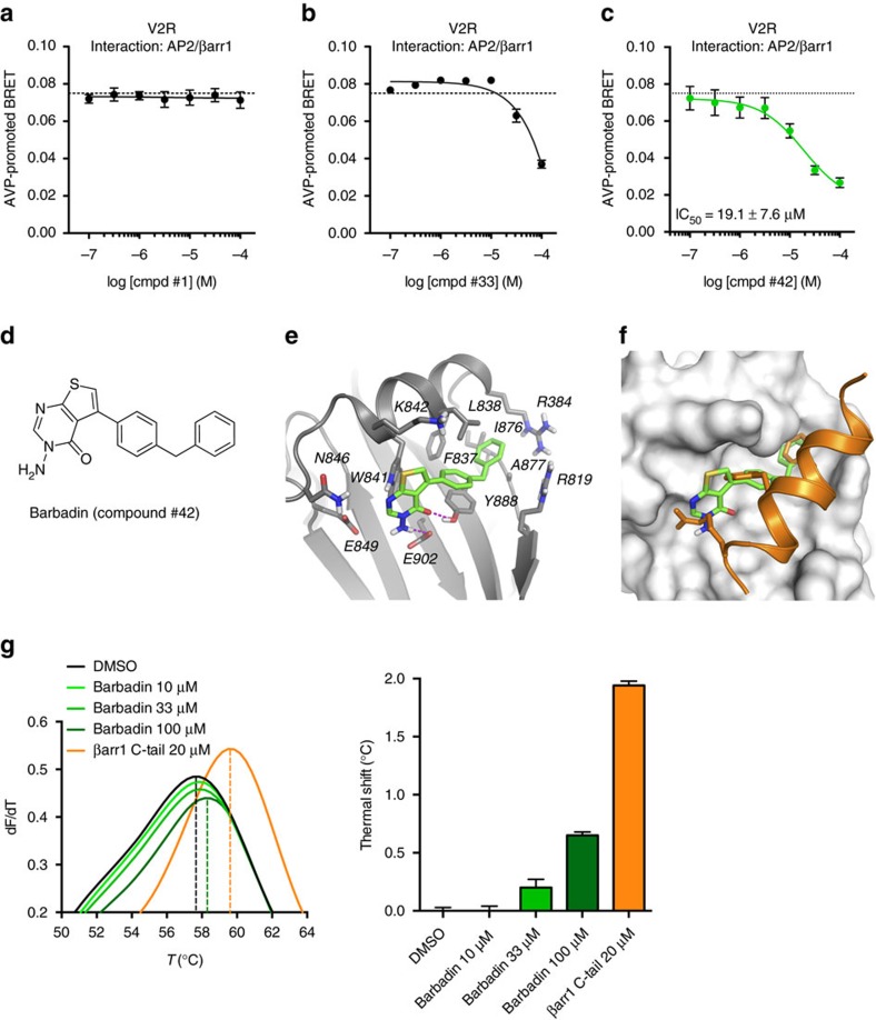
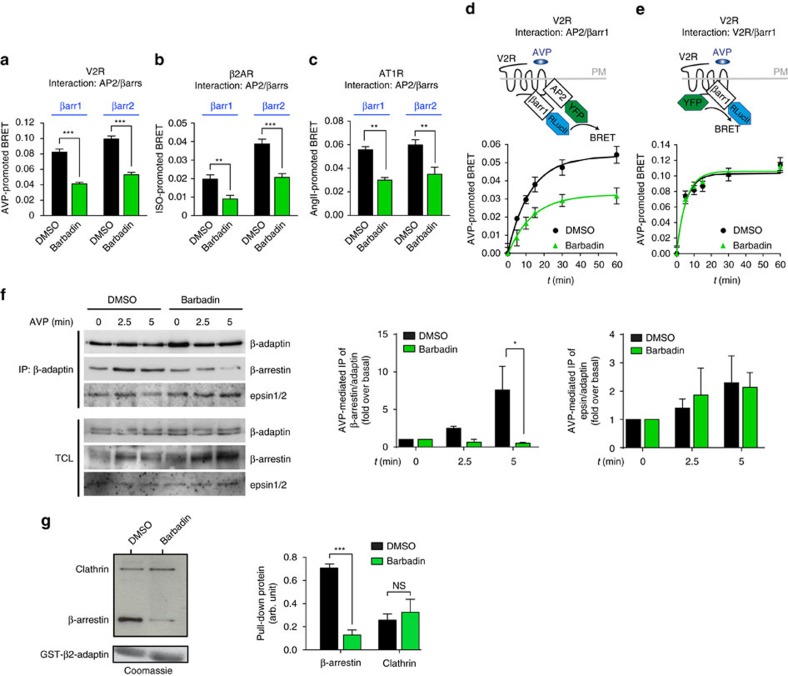
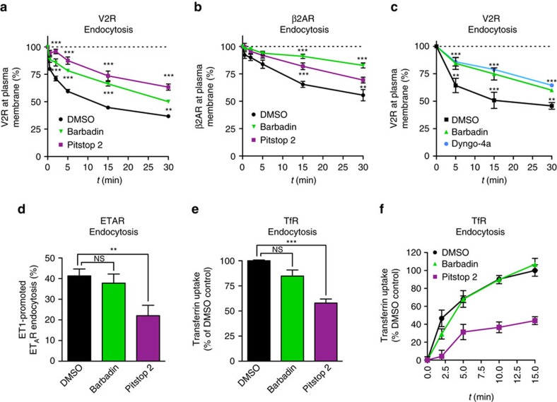
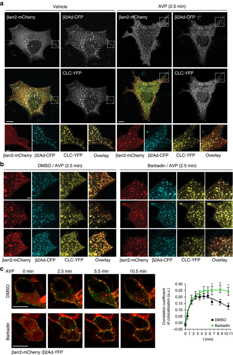
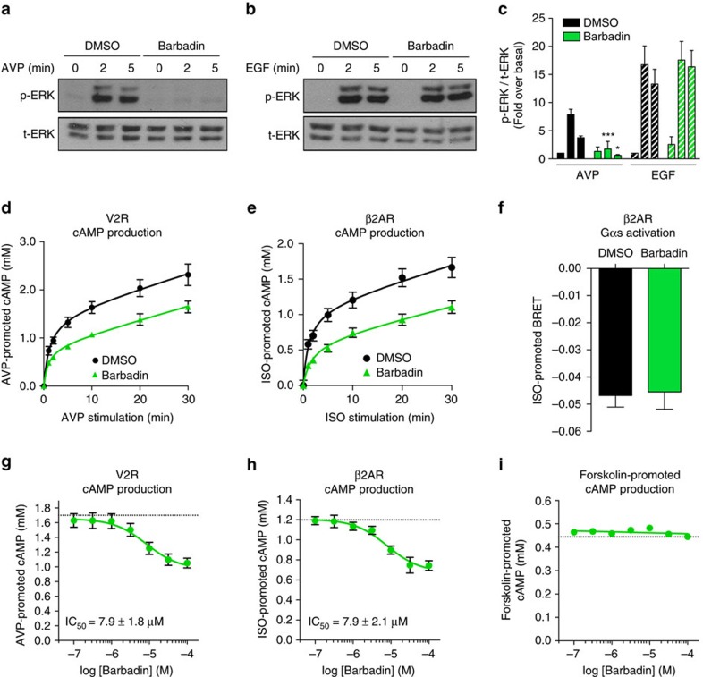
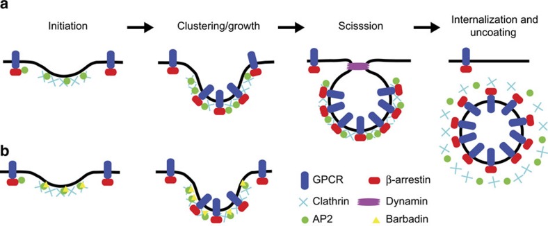
In addition to G protein-coupled receptor (GPCR) desensitization and endocytosis, β-arrestin recruitment to ligand-stimulated GPCRs promotes non-canonical signalling cascades. Distinguishing the respective contributions of β-arrestin recruitment to the receptor and β-arrestin-promoted endocytosis in propagating receptor signalling has been limited by the lack of selective analytical tools. Here, using a combination of virtual screening and cell-based assays, we have identified a small molecule that selectively inhibits the interaction between β-arrestin and the β2-adaptin subunit of the clathrin adaptor protein AP2 without interfering with the formation of receptor/β-arrestin complexes. This selective β-arrestin/β2-adaptin inhibitor (Barbadin) blocks agonist-promoted endocytosis of the prototypical β2-adrenergic (β2AR), V2-vasopressin (V2R) and angiotensin-II type-1 (AT1R) receptors, but does not affect β-arrestin-independent (transferrin) or AP2-independent (endothelin-A) receptor internalization. Interestingly, Barbadin fully blocks V2R-stimulated ERK1/2 activation and blunts cAMP accumulation promoted by both V2R and β2AR, supporting the concept of β-arrestin/AP2-dependent signalling for both G protein-dependent and -independent pathways.


Conflict of interest statement

The authors declare no competing financial interests.

Figures

Figure 1
Figure 1. Screen for inhibitors of the interaction between β2-adaptin and β-arrestin.
(a) Flowchart outlining the steps of the virtual screening and the selection of candidate molecules to be tested. (b) Schematic representation and dynamic range of the BRET-based assay used to test the compounds selected from the virtual screen that monitors the β2-adaptin/β-arrestin1 interaction. HEK293T cells were transfected with β2-adaptin-YFP, β-arrestin1-RlucII and myc-V2R. BRET was measured following V2R stimulation with 100 nM AVP for 45 min. Data are the mean±s.e.m. of three independent experiments. Statistical analysis was performed using a paired Student's test (***P<0.001). (c) β2-adaptin/β-arrestin1 interaction was assessed as described in (b) with AVP-stimulation following pre-incubation with the indicated compounds (100 μM) for 25 min. Data are the mean±s.e.m. of three independent experiments. One-way ANOVA followed by Tuckey's post-hoc tests were used to assess the statistical significance of the compound-induced BRET modulation compared to DMSO (***P<0.001).
Figure 2
Figure 2. Identification of Barbadin (compound #42) as an inhibitor of the interaction between β2-adaptin and β-arrestin.
(ac) Concentration-response curves of compounds #1, #33 and #42 selected from the BRET-based screen (Fig. 1) using the same β2-adaptin/β-arrestin1 interaction assay. Dotted line represents the level of AVP-promoted BRET upon pre-incubation with DMSO. Data are the mean±s.e.m. of three independent experiments. (d) Chemical structure of Barbadin (IUPAC name: 3-amino-5-(4-benzylphenyl)-3H,4H-thieno[2,3-d]pyrimidin-4-one). (e,f) Docking pose of Barbadin (green sticks) within the groove of β2-adaptin platform subdomain (grey ribbon (e), or surface (f)) that is the site of interaction with β-arrestin (orange ribbon and sticks (f)). β2-adaptin residues known to interact with β-arrestin are labelled and shown as grey stick. Hydrogen-bonds interactions between Barbadin and both Tyr-888 and Glu-902 are depicted as magenta dotted lines (e). Superimposition of Barbadin with the β-arrestin1 C-terminus peptide as in the co-crystal structure (PDB entry 2IV8), where Phe-388, Phe-391 and Arg-395 are the three key residues for β-arrestin binding interaction (shown from top to bottom as orange sticks) (f). (g) Thermal denaturation of β2-adaptin (residues 700–937) and concentration-dependent stabilization effect of Barbadin. The maximum of the first derivatives of fluorescence (dF/dT) data from differential scanning fluorimetry corresponds to the melting temperatures (Tm, indicated with a dotted line) of β2-adaptin in the presence of DMSO, Barbadin or β-arrestin1 C-tail peptide (positive control) at the indicated concentrations.
Figure 3
Figure 3. Barbadin specifically blocks the interaction between β2-adaptin and β-arrestin.
(ac) BRET-based assay monitoring the interaction between β2-adaptin-YFP and either β-arrestin1-RlucII or β-arrestin2-RlucII. HEK293T cells were pre-incubated with DMSO or Barbadin (100 μM) for 30 min before 45 min receptor stimulation with AVP (100 nM, a), ISO (10 μM, b) or AngII (100 nM, c). Data are the mean±s.e.m. of at least three independent experiments and unpaired t-test were used to assess statistical significance (***P<0.001; **P<0.005). (d,e) BRET-based kinetics monitoring the interaction between β-arrestin1-RlucII and β2-adaptin-YFP (d) or V2R-YFP (e) in HEK293T cells pretreated with DMSO or Barbadin (100 μM) for 30 min before receptor stimulation with AVP (100 nM) for the indicated times. Data are the mean±s.e.m. of three independent experiments. (f) Effect of Barbadin on the co-immunoprecipitation between β-arrestins and AP2. HEK293SL cells expressing V2R and Flag-β-arrestin2 were pretreated for 20 min with DMSO or Barbadin (50 μM) before stimulation by AVP (1 μM) for 2.5 or 5 min. Endogenous AP2 complexes (using the AP1/2 antibody) were immunoprecipitated (IP) from total cell lysates (TCL), and then analysed by western blot using anti-Flag, anti-epsin or anti-adaptin antibodies as described in the Material and Methods. TCLs represent 5% of input used for IP. IP were quantified over three independent experiments and statistical significance was assessed by a two-way ANOVA followed by Bonferroni's post-hoc tests (*P<0.05). (g) Effects of Barbadin on the pull-down of β-arrestin1 and clathrin with GST-β2-adaptin. DMSO or Barbadin (100 μM) were incubated with GST-β2-adaptin (592–937) beads. HEK293T cells transfected with Flag-β-arrestin1 were lysed and added to the beads. The amounts of GST-β2-adaptin were detected by Coomassie, whereas β-arrestin1 and clathrin associated with GST-β2-adaptin were detected by western blot using anti-Flag and anti-clathrin (heavy chain) antibodies, respectively. Relative intensities were normalized to the GST input for each condition and densitometry data are the mean±s.e.m. of three independent experiments and analysed using a two-way ANOVA followed by Bonferroni's post-hoc tests (NS, non-significant; ***P<0.001).
Figure 4
Figure 4. Barbadin inhibits GPCRs endocytosis.
(a,e) Schematic representation of the ebBRET-based assay used to follow agonist-induced receptor loss from the cell surface by monitoring the interaction between receptor-RLucII and rGFP-CAAX (a) or its translocation into endosomes using rGFP-FYVE (e). (bd,fh) V2R, β2AR or AT1R interaction with either rGFP-CAAX (bd, respectively) or rGFP-FYVE (fh, respectively) was assessed by BRET following HEK293T cells pre-incubation with DMSO or Barbadin (100 μM) for 30 min before AVP (100 nM), ISO (10 μM) or AngII (1 μM) stimulation for the indicated times. Data are the mean±s.e.m. of a least three independent experiments. (i) V2R localization was imaged by BRET. HEK293T cells were transfected with V2R-RlucII and rGFP-CAAX, pretreated with DMSO or Barbadin (100 μM) for 30 min and then stimulated with 100 nM AVP for 30 min. To generate BRET images, the ratio of acceptor photon counts to donor photon counts was calculated for each pixel and expressed as a colour-coded heat map (lowest being black and purple, and highest red and white). Scale bar, 10 μm.
Figure 5
Figure 5. Barbadin inhibits the β-arrestin- and AP2-dependent endocytosis of GPCRs.
(ac) Cell surface expression of HA-V2R-Venus (a,c) or HA-β2AR-Venus (b) transfected in HEK293T cells was monitored by FACS following pre-incubation with DMSO, Barbadin (100 μM), Pitstop2 (100 μM) or Dyngo-4a (30 μM) for 30 min, before agonist stimulation (AVP (100 nM, a,c), ISO (10 μM, b)) at the indicated times. Data are the mean±s.e.m. of at least three independent experiments. Statistical significance of the effect of Barbadin, Pitstop 2 and Dyngo, as compared to DMSO, was assessed by a two-way ANOVA followed by Bonferroni's post-hoc tests (**P<0.01; ***P<0.001). (d) Cell surface expression of HA-ETAR was monitored by FACS following pre-incubation with DMSO, Barbadin (100 μM) or Pitstop2 (100 μM) for 30 min, before ET1 (10 nM) stimulation for 30 min. Data are the mean±s.e.m. of at least three independent experiments. Statistical significance was assessed by a one-way ANOVA followed by Tuckey's post-hoc tests (NS, non-significant; **P<0.01). (e,f) Native transferrin receptor (TfR) uptake was monitored by FACS in HeLa cells. Cells were pretreated with DMSO, Barbadin (100 μM), or Pitstop2 (100 μM) for 30 min, then incubated with Alexa Fluor 633-conjugated transferrin antibody (100 μg ml−1) for 30 min at 4 °C, and finally shifted to 37 °C for 15 min (e) or the indicated time (f). Data are the mean±s.e.m. of three independent experiments. Statistical significance was assessed by a one-way ANOVA followed by Tuckey's post-hoc tests (NS, non-significant; **P<0.01; ***P<0.001).
Figure 6
Figure 6. Barbadin induces the retention of receptor/β-arrestin complexes in clathrin-coated pits (CCPs) at the membrane.
(a,b) Confocal images and colocalization of β-arrestin2, β2-adaptin, and clathrin light chain-(CLC) in CCPs from AVP-stimulated cells expressing V2R. β2-adaptin depleted HEK293 cells (CRISPR-β2Ad-LY5) were transfected with β-arrestin2-mCherry, β2-adaptin-CFP and CLC-YFP, and HA-V2R, and serum-starved for 30 min in the absence or presence of Barbadin (10 μM), before being either left non-stimulated (vehicle) or stimulated with AVP (1 μM) for 2.5 min. Cells were then fixed as described in the Methods section before visualization. Shown in the top panels are black and white micrographs of acquired fluorescent signals from the three-tagged proteins in each channel (red, cyan and yellow), and in colour, are the overlay images. Lower colour insets are close-up images from boxed areas. (b) Colour images represent individual and overlay fluorescent signals taken from different areas, from cells transfected, treated and stimulated as in a. Scale bars in images of whole cells, 10 μm; and insets, 2 μm. Colocalization quantification is presented in Supplementary Fig. 7b–c. (c) Colocalization and quantification of β-arrestin2-mCherry and β2-adaptin-YFP in live HEK293T cells stably expressing V2R. Cells were pretreated with DMSO or Barbadin (10 μM) for 30 min before stimulation with AVP (1 μM) for the indicated durations. Co-localization was quantified using the Pearson correlation coefficient over three independent experiments using 33 or 24 cells for DMSO and Barbadin condition, respectively. Statistical significance of the effect of Barbadin as compared to DMSO was assessed by a two-way ANOVA followed by Bonferroni's post-hoc tests (*P<0.05).
Figure 7
Figure 7. Barbadin inhibits ERK1/2 activation and cAMP accumulation following agonist-stimulation of GPCR.
(ac) Kinetics of ERK1/2 phosphorylation in HEK293T cells expressing V2R (a) or EGFR (b) and pretreated with DMSO or Barbadin (50 μM) for 30 min before stimulation with AVP (100 nM, a) or EGF (100 ng ml−1, b) at the indicated times. Western blots were quantified (c) and data shown are the mean±s.e.m. of four independent experiments and analysed using a two-way ANOVA followed by Bonferroni's post-tests (*P<0.05; ***P<0.001). (d,e) Kinetics of the agonist-promoted accumulation of cAMP in HEK293T cells stably expressing V2R (d) or β2AR (e) and pretreated with DMSO or Barbadin (50 μM) for 30 min before stimulation with AVP (100 nM, d) or ISO (10 μM, e) at the indicated times. Data are the mean±s.e.m. of three independent experiments. (f) ISO-promoted Gs activation measured by BRET in HEK293T cells transfected with HA-β2AR, Gαs-RLucII, Gβ1 and Gγ2-GFP10, pretreated with DMSO or Barbadin (100 μM) for 30 min, before ISO (10 μM) stimulation for 5 min. (g,h) Concentration-response curves of Barbadin effect on the intracellular cAMP production, in HEK293T cells stably expressing the V2R (g) or the β2AR (h), upon agonist stimulation for 15 min with AVP (100 nM) or ISO (1 μM), respectively, following pre-treatment with DMSO (grey dotted line) or Barbadin (green) at the indicated concentrations for 30 min. Data are the mean±s.e.m. of three independent experiments. (i) Concentration-response curves of Barbadin effect on the intracellular cAMP production, in HEK293T cells stimulated with forskolin (10 μM) for 15 min. Same DMSO and Barbadin pre-treatments as in (g,h). Data are the mean±s.e.m. of three independent experiments.
Figure 8
Figure 8. Model for the biogenesis of clathrin-coated pits (CCPs) and Barbadin's effect on GPCRs endocytosis.
(a) Initiation of CCPs formation involves the association of AP2 with phosphatidylinositol 4,5-bisphosphate (PtdIns(4,5)P2) at the plasma membrane (PM) and the recruitment of clathrin to AP2 at sites of nucleation. Growth and stabilization of coated-vesicles requires the recruitment of additional AP2 and clathrin, and endocytic accessory proteins like epsin (not shown here) and β-arrestins. Agonist-activated GPCR/β-arrestin complexes accumulate in CCPs through β-arrestin's interactions with AP2 (β2 and μ subunits) and clathrin, stabilizing further the growing coated-vesicles. Stabilization of GPCR/β-arrestin complexes and the recruitment of endocytic effectors like dynamin allows the matured coated-vesicles to commit for invagination and scission from the PM, which is followed by the uncoating (that is, release of AP2 and clathrin) of internalizing vesicles. (b) In the presence of Barbadin, initiation of CCPs still occurs because clathrin and AP2 are effectively recruited at sites of nucleation. Some receptor/β-arrestin complexes will still coalesce into nucleating CCPs through β-arrestin's binding with clathrin and its low affinity interaction to AP2 (for example, via the μ subunit). Because Barbadin prevents the interactions of β-arrestin with the appendage domain on the β2-subunit of AP2, which prevents the stable formation of sufficient GPCR/β-arrestin complexes in CCPs, the maturation of the coated-vesicles is hampered, and receptor internalization impeded.

References

    1. Sorkin A. & von Zastrow M. Endocytosis and signalling: intertwining molecular networks. Nat. Rev. Mol. Cell Biol. 10, 609–622 (2009). - PMC - PubMed
    1. Shenoy S. K. & Lefkowitz R. J. Beta-Arrestin-mediated receptor trafficking and signal transduction. Trends Pharmacol. Sci. 32, 521–533 (2011). - PMC - PubMed
    1. Lohse M. J., Benovic J. L., Codina J., Caron M. G. & Lefkowitz R. J. Beta-Arrestin: a protein that regulates beta-adrenergic receptor function. Science 248, 1547–1550 (1990). - PubMed
    1. Ferguson S. S. et al. Role of beta-arrestin in mediating agonist-promoted G protein-coupled receptor internalization. Science 271, 363–366 (1996). - PubMed
    1. Goodman O. B. Jr et al. Beta-arrestin acts as a clathrin adaptor in endocytosis of the beta2-adrenergic receptor. Nature 383, 447–450 (1996). - PubMed

Publication types

MeSH terms

Substances