. 2018 May;23(5):1336-1344.
doi: 10.1038/mp.2017.56. Epub 2017 Apr 18.

A simplified protocol for differentiation of electrophysiologically mature neuronal networks from human induced pluripotent stem cells

Affiliations

A simplified protocol for differentiation of electrophysiologically mature neuronal networks from human induced pluripotent stem cells

N Gunhanlar et al. Mol Psychiatry. 2018 May.

Abstract

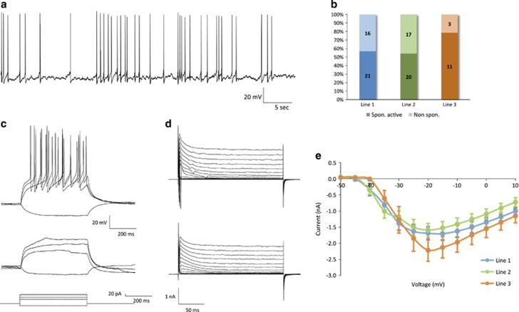
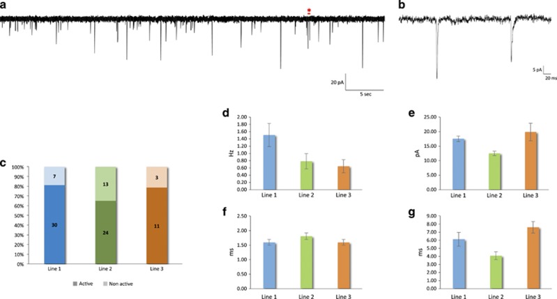
Progress in elucidating the molecular and cellular pathophysiology of neuropsychiatric disorders has been hindered by the limited availability of living human brain tissue. The emergence of induced pluripotent stem cells (iPSCs) has offered a unique alternative strategy using patient-derived functional neuronal networks. However, methods for reliably generating iPSC-derived neurons with mature electrophysiological characteristics have been difficult to develop. Here, we report a simplified differentiation protocol that yields electrophysiologically mature iPSC-derived cortical lineage neuronal networks without the need for astrocyte co-culture or specialized media. This protocol generates a consistent 60:40 ratio of neurons and astrocytes that arise from a common forebrain neural progenitor. Whole-cell patch-clamp recordings of 114 neurons derived from three independent iPSC lines confirmed their electrophysiological maturity, including resting membrane potential (-58.2±1.0 mV), capacitance (49.1±2.9 pF), action potential (AP) threshold (-50.9±0.5 mV) and AP amplitude (66.5±1.3 mV). Nearly 100% of neurons were capable of firing APs, of which 79% had sustained trains of mature APs with minimal accommodation (peak AP frequency: 11.9±0.5 Hz) and 74% exhibited spontaneous synaptic activity (amplitude, 16.03±0.82 pA; frequency, 1.09±0.17 Hz). We expect this protocol to be of broad applicability for implementing iPSC-based neuronal network models of neuropsychiatric disorders.


Conflict of interest statement

The authors declare no conflict of interest.

Figures

Figure 1
Figure 1
Generation and characterization of NPCs and neuronal networks from iPSCs. (a) Scheme illustrating the major developmental stages of the protocol for generating NPCs and neuronal networks. (b) Immunostaining for NPC markers Nestin, SOX2, Vimentin and FOXG1 (scale bars=30 μm). (c) Proportion of NeuN+ and GFAP+ cells (days 56–70). (d) Immunostaining for glial marker GFAP, and mature neuronal markers MAP2 and NeuN (top, scale bar=20 μm; bottom, scale bar=10 μm). (e) Co-labeling of pre- and postsynaptic marker proteins, Synapsin and PSD95 (scale bar=2 μm). (f) Quantification of Synapsin+, PSD95+ and double-labeled puncta density (n=20 neurons). EB, embryoid body; GFAP, glial fibrillary acidic protein; iPSC, induced pluripotent stem cells; NPC, neural precursor cells.
Figure 2
Figure 2
Cortical layer markers in neuronal networks. Cultures were stained at day 56 following the initiation of NPC differentiation for (a) BRN2 marker of late cortical progenitors and upper layer (II-IV) neurons, and mature dendritic marker MAP2, (b) TBR1 that is expressed by deep layer neurons (V and VI) and in the subplate, (c) FOXP2 expressed in deep layer (V and VI) neurons, (d) CUX1 marker of upper layer (II–IV) neurons and telencephalic marker FOXG1 and (e) CUX2 marker of upper layer (II–IV) neurons and SATB2 expressed in corticocortical projection neurons from layer V and upper layers. (f) CTIP2 expression in deep layer glutamatergic projection neurons. NPC, neural precursor cells.
Figure 3
Figure 3
Active and passive electrophysiological properties. (a) Representative traces from a neuron firing repetitive mature APs during depolarizing constant-current injections. Current steps are shown in the bottom panel (Vm=−75 mV). The lowest depolarizing step indicates the minimal current needed to evoke an action potential, and the highest step corresponds to the current at which the response frequency became saturated. (b) Percentage of repetitive versus nonrepetitively firing neurons. (c) Frequency–current (F-I) plot among repetitively firing neurons. (d–k) Active and passive membrane properties. AP parameters were calculated from the first evoked spike. (d) Input resistance (F=3.65, P=0.03), (e) resting membrane potential (F=0.82, P=0.44), (f) capacitance (F=0.18, P=0.84), (g) AP threshold (F=1.25, P=0.29), (h) AP amplitude (F=1.01, P=0.37), (i) AP half-width (F=4.70, P=0.012), (j) AP rise time (F=1.23, P=0.30) and (k) decay time (F=4.62, P=0.013). AP, action potential.
Figure 4
Figure 4
Spontaneous action potentials. (a) Representative current-clamp recording from a spontaneously active neuron (Vm=−68 mV). (b) Percentage of neurons with spontaneous AP firing. (c) Voltage responses of the same neuron in (a) to hyperpolarizing or depolarizing current injections (bottom panel), before (top panel) and after (middle panel) TTX application (Vm=−75 mV). (d) Sodium currents were abolished by TTX (before, top panel; after, bottom panel) (Vm=−80 mV). (e) Voltage dependence of the peak amplitude of the sodium current.
Figure 5
Figure 5
Neuronal network synaptic activity. (a) Representative voltage-clamp recording from a neuron with spontaneous synaptic input (Vm=−80 mV). (b) Zoom-in of the region in (a) marked by the red asterisk, containing two postsynaptic events. (c) Percentage of neurons exhibiting spontaneous synaptic input. (d–g) Spontaneous postsynaptic currents: (d) frequency (F=2.55, P=0.09), (e) amplitude (F=7.25, P=0.001; post hoc Tukey: P=0.01 for line 1 vs 2, P=0.004 for line 2 vs 3 and P=0.52 for line 1 vs 3), (f) rise time (F=1.24, P=0.30) and (g) decay time (P=0.023, F=4.01).

References

    1. Sekar A, Bialas AR, de Rivera H, Davis A, Hammond TR, Kamitaki N et al. Schizophrenia risk from complex variation of complement component 4. Nature 2016; 530: 177–183. - PMC - PubMed
    1. Sztainberg Y, Chen H, Swann JW, Hao S, Tang B, Wu Z et al. Reversal of phenotypes in MECP2 duplication mice using genetic rescue or antisense oligonucleotides. Nature 2015; 528: 123–126. - PMC - PubMed
    1. Willem M, Tahirovic S, Busche MA, Ovsepian SV, Chafai M, Kootar S et al. η-Secretase processing of APP inhibits neuronal activity in the hippocampus. Nature 2015; 526: 443–447. - PMC - PubMed
    1. Cirulli ET, Lasseigne BN, Petrovski S, Sapp PC, Dion PA, Leblond CS et al. Exome sequencing in amyotrophic lateral sclerosis identifies risk genes and pathways. Science 2015; 347: 1436–1441. - PMC - PubMed
    1. Meng L, Ward AJ, Chun S, Bennett CF, Beaudet AL, Rigo F. Towards a therapy for Angelman syndrome by targeting a long non-coding RNA. Nature 2014; 518: 409–412. - PMC - PubMed

Publication types

MeSH terms