. 2017 Jan 31:3:17003.
doi: 10.1038/celldisc.2017.3. eCollection 2017.

Chemical biology reveals CARF as a positive regulator of canonical Wnt signaling by promoting TCF/β-catenin transcriptional activity

Affiliations

Chemical biology reveals CARF as a positive regulator of canonical Wnt signaling by promoting TCF/β-catenin transcriptional activity

Xiaoli He et al. Cell Discov. .

Abstract

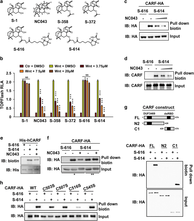
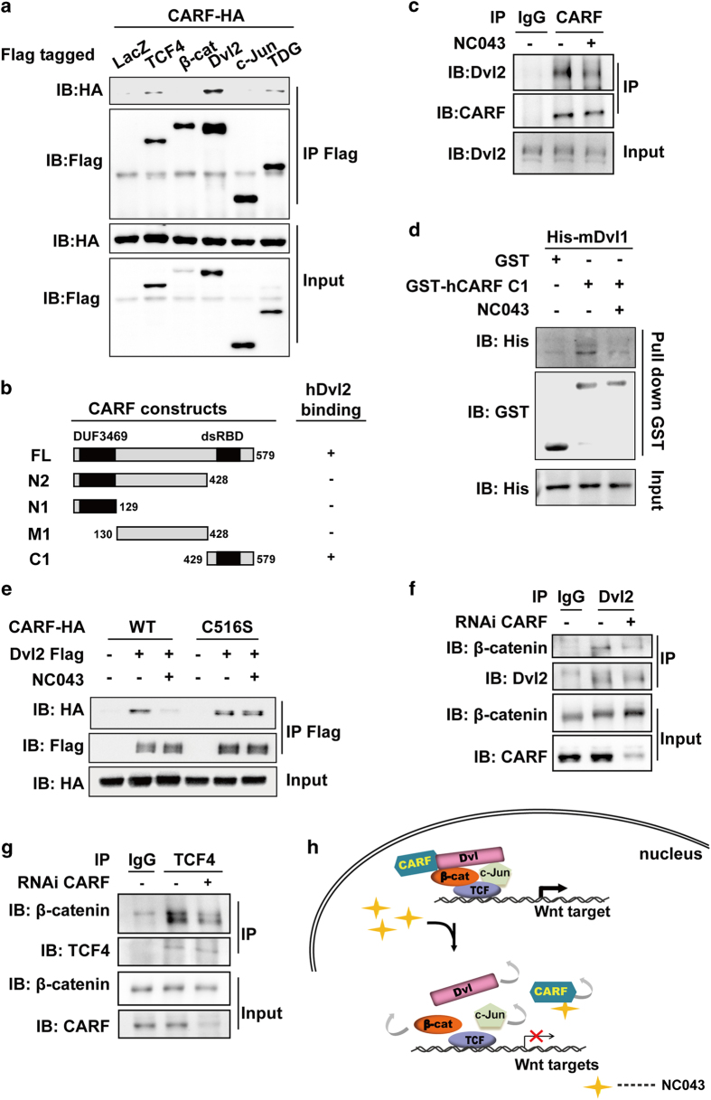
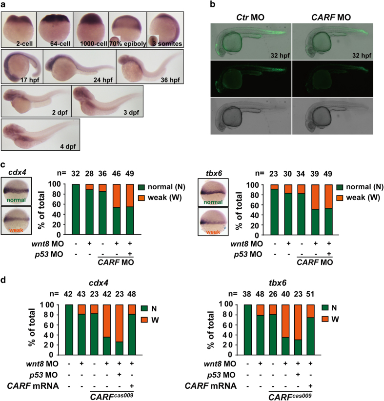
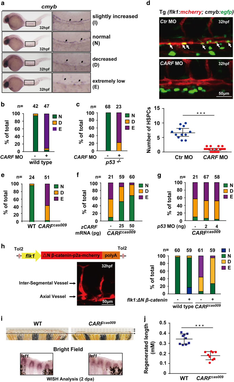
Wnt/β-catenin signaling regulates multiple biological processes and aberration of this pathway is frequently observed in human cancers. Previously, we uncovered NC043 as a small-molecule inhibitor of Wnt/β-catenin signaling. Here, we identified CARF as the cellular target of NC043. We found that NC043 binds directly to CARF through forming a covalent bond with the Cys-516 residue of CARF. Further study revealed that CARF interacts with Dvl, which potentiates the Dvl-c-Jun-β-catenin-TCF transcriptional complex and thus promotes Wnt signaling activation. NC043 could disrupt the interaction between CARF and Dvl, thereby impairing Wnt signal transduction. In line with this, knockdown of CARF in zebrafish leads to impairment of embryonic development, hematopoietic stem cell generation and caudal fin regeneration. Collectively, we identified CARF as the cellular target of NC043 and revealed CARF as a positive regulator of Wnt/β-catenin signal transduction.

Keywords: CARF; NC043; Wnt signaling; chemical biology; small molecule.


Conflict of interest statement

The authors declare no conflict of interest.

Figures

Figure 1
Figure 1
CARF is a target of NC043. (a) Chemical structures of NC043 and its analogs. (b) Effects of NC043 and its analogs on TOPFlash reporter activity. After 18 h of transfection, HEK293 cells were treated with the control (Ctr) conditioned medium (CM) or the Wnt3a CM plus the indicated small molecules for an additional 6 h before the luciferase activity assays. RLA, relative luciferase activity. Error bars indicate the s.d. of triplicate assays in one experiment. Each experiment was repeated at least three times. (*P-value<0.05; **P-value<0.01; ***P-value<0.001). (c) S-614 binds to CARF overexpressed in HEK293T cells. HEK293T overexpressed HA-tagged CARF were treated with 7.5 μm S-616 or S-614 plus with 0, 3.75 and 15 μm of NC043 for 1 h. The cell lysates were used for biotin-pulldown assay. The levels of CARF before and after pulldown were detected through western blotting analysis with HA antibody. (d) S-614 binds to endogenous CARF. After 1 h of 7.5 μm S-616 or S-614 plus with 0, 3.75 and 15 μm NC043 treatment, HEK293 cells were lysed, and followed by biotin-pulldown and western blotting analysis using a CARF specific antibody (anti-CARF). S-616 was used as a negative control. Error bars indicate the s.d. of triplicate assays in one experiment. Each experiment was repeated at least three times. (e) S-614 directly binds to purified CARF expressed in bacteria. The lysate of His6-tagged hCARF expressed E.coli. was incubated with Ni-beads for 1 h. After three times of wash, the beads were resuspended and incubated with small compounds (7.5 μm S-614 or S-616 and 15 μm NC043) as indicated for 1 h at 4 °C. Samples were subjected to biotin and His detaction. (f) NC043 binds to CARF covalently. After transfection, HEK293T cells were treated with 7.5 μm S-614 or S-616 and 15 μm NC043 as indicated, followed by biotin-pulldown and western blotting analysis. ‘*’ Cells were incubated in DMSO-contained medium for half-an-hour, and then treated with S-616/DMSO, S-614/DMSO or S-614/NC043 (lane 1-3) for 1 h; ‘**’ After S-614 pre-treatment, cells were incubated in S-614/DMSO or S-614/NC043 (lane 4–5) supplemented medium for additional 1 h. (g) Mapping the region on CARF responsible for binding NC043. Upper panel: schematic representation of full-length CARF (FL) and its truncations. After plasmids transfection and compounds incubation, HEK293T cells were used for biotin-pulldown and western blotting analysis. (h) C516 is the critical site for CARF to bind NC043. HEK293T cells expressed CARF (WT) and the indicated mutants were used for S614 binding assay followed by western blotting analysis.
Figure 2
Figure 2
CARF promotes canonical Wnt signaling downstream of β-catenin accumulation. (a) Overexpressed CARF promotes Wnt signaling. After transfection as indicated, HEK293 cells were used for TOPFlash reporter assay. (b) Knockdown of CARF represses Wnt3a-induced TOPFlash activity. After CARF knockdown with the indicated siRNAs for 48 h, HEK293 cells were subjected to reporter assay. (c) Expression of Axin2 was decreased by CARF knockdown and restored by CARF overexpression. Inner panel shows the expression levels of CARF with β-actin as the loading control. ‘CARF*’ indicates that the plasmid is a siRNA-resistant one. (d) Expression of Wnt target genes was inhibited by CARF knockdown in Saos-2 cells. After 42 h of CARF RNAi and additional 6 h of Wnt CM treatment, cells were subjected to RT-PCR. (e, f) Profile of genes regulated by Wnt, β-catenin and CARF. (e) Venn diagram for the relationship between CARF and β-catenin regulated genes among Wnt-responded ones. A, Set of genes responding to Wnt3a; B, Set of genes with a decreased expression after CARF knockdown; C, Set of genes downregulated by β-catenin knockdown; D, The intersection of B&C. (f) Expression pattern of the 40 genes from the set D described in e. All the analyses were based on two independent experiments. (g) Knockdown of CARF repressed expression of the Wnt target genes in SW480. After 48 h of CARF RNAi, cells were subjected to RT-PCR. Error bars indicate the s.d. of triplicate assays in one experiment. Each experiment was repeated at least three times.
Figure 3
Figure 3
CARF facilitates assemble of the multi-components transcriptional complex through interacting with Dvl. (a) CARF interacts with Dvl2. HEK293T cells were transfected as indicated, and followed by immunoprecipitation (IP) using anti-Flag and western blotting analysis. (b) Schematic representation of the full-length and truncated CARF with the indicated binding affinity for Dvl2. (c) CARF interacts with Dvl2 in vivo and this interaction is disrupted by NC043. Nuclear extract of HEK293 cells with or without NC043 treatment were subjected to IP using anti-CARF followed by immunoblotting. IgG was used as a negative control for the IP analysis. (d) NC043 disrupts CARF-Dvl2 interaction in vitro. GST-hCARF and His-mDvl1 expressed in E.coli were used for GST-pulldown with or without NC043 incubation, followed by western blotting analysis. (e) NC043 disrupts the interaction of Dvl2 with WT CARF but not with C516S mutant. HEK293T cells transfected with indicated plasmids were subjected to IP analysis using anti-Flag followed by western blotting analysis using anti-HA. (f, g) CARF knockdown decreases Dvl–β-catenin association (f) and TCF4–β-catenin association (g). The nuclear extracts from CARF-deprived HEK293T cells were subjected to IP using anti-Dvl2 and anti-TCF4 followed by western blotting analysis. (h) A model for how CARF facilitates TCF transcriptional activity and NC043 inhibits Wnt signaling. On Wnt stimulation, CARF facilitates assembly of the Dvl–c-Jun–β-catenin–TCF complex through interacting directly with Dvl. When NC043 is present, NC043 binds to CARF covalently via targeting its C516 residue, which disrupts CARF–Dvl interaction and consequently impairs the TCF multi-components complex.
Figure 4
Figure 4
CARF facilitates Wnt/β-catenin signaling in zebrafish embryogenesis. (a) The whole mount in situ analysis of CARF expression in zebrafish embryogenesis. The embryos at indicated developmental stages were fixed for in situ hybridization with CARF probe. (b) CARF knockdown attenuates Wnt activation in Tg(tcf:egfp) line. (c, d) CARF cas009 mutant or CARF morphants show reduced Wnt signaling activity via a p53-independet manner. Embryos injected with the indicated morpholinos or morpholino-mRNA mixture were fixed at the 60% epiboly stage and then analyzed by WISH with cdx4 (left) or tbx6 (right) probe. The relative expression of each marker was classified into two categories: normal (N) or weak (W). The number of the total embryos scored (n) is shown on the top of each bar.
Figure 5
Figure 5
Loss of CARF attenuates HSPC formation and caudal fin regeneration. (a–d) Knockdown CARF limits HSPC formation. Zebrafish embryos at 32 hpf were fixed for in situ hybridization with cmyb probe, and then classified into three categories: (a) Representative image for slightly increased (I), normal (N), decreased (D) or extremely low (E) level of c-myb WISH analysis. Knockdown CARF dramatically inhibits HSPC formation (b) which could not be rescued via p53 MO co-injection (c) and further validated by the live image of HSPC budding events in AGM of zebrafish Tg(flk1:mcherry;cmyb:egfp) line (d). (e–g) CARF cas009 mutant exhibits reduced HSPC formation via a p53-independent manner. Injection of zebrafish CARF (zCARF) mRNA (f) but not p53 MO (g) restores decreased cmyb expression in CARF cas009 mutants (e). (h) Schematic diagram of Tol2 transposase-mediated transient transgenesis of endothelial-specific promoter (flk1)-derived expression of constitutive activated β-catenin (ΔN β-catenin) and mCherry chimera protein in zebrafish embryos, which rescues hematopoietic defects in CARF cas009 mutants while slightly increases HSC formation in wildtype zebrafish. (i, j) Delayed fin regeneration of CARF cas009 mutants. Adult CARF cas009 zebrafish were executed caudal fin amputation and then cultured for regeneration. The regenerated sections were cut down at 2 dpa for either WISH analysis with lef1 probes or length measurement.

References

    1. Clevers H . Wnt/beta-catenin signaling in development and disease. Cell 2006; 127: 469–480. - PubMed
    1. Klaus A , Birchmeier W . Wnt signalling and its impact on development and cancer. Nat Rev Cancer 2008; 8: 387–398. - PubMed
    1. Wang W , Liu H , Wang S , Hao X , Li L . A diterpenoid derivative 15-oxospiramilactone inhibits Wnt/beta-catenin signaling and colon cancer cell tumorigenesis. Cell Res 2011; 21: 730–740. - PMC - PubMed
    1. Wang S , Yin J , Chen D et al. Small-molecule modulation of Wnt signaling via modulating the Axin-LRP5/6 interaction. Nat Chem Biol 2013; 9: 579–585. - PubMed
    1. Chen B , Dodge ME , Tang W et al. Small molecule-mediated disruption of Wnt-dependent signaling in tissue regeneration and cancer. Nat Chem Biol 2009; 5: 100–107. - PMC - PubMed