

Giant Ureteral Fibroepithelial Polyp with Intermittent Prolapse Reaching the Urethral Meatus: A Case Report
- PMID: 28417075
- PMCID: PMC5388910
- DOI: 10.1016/j.eucr.2017.03.015
Giant Ureteral Fibroepithelial Polyp with Intermittent Prolapse Reaching the Urethral Meatus: A Case Report
Abstract
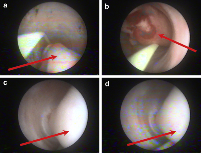
Ureteral fibroepithelial polyps (UFPs) are rare non-epithelial benign tumors of the urinary tract. Treatment of such cases ranges from conservative management to surgical resection of the polyp. Hereby, we present a rare case of a 37-year-old female patient with giant 14 cm UFP of the distal left ureter, successfully resected by ureteroscopic electrocauterization. Several cases of UFPs have been previously reported in world literature describing polyps extending into the bladder; yet, our case is the first to present a giant UFP that extends beyond the bladder cavity protruding outside the urethral meatus as a red fleshy mass.
Keywords: Case report; Cystoureteroscopy; Fibroepithelial polyp; Ureteral polyp.
Figures


References
-
- Bolton D., Stoller M.L., Irby P., III Fibroepithelial ureteral polyps and urolithiasis. Urology. 1994;44(4):582–587. - PubMed
-
- Uġras S., Odabas Ö., Aydin S., Yilmaz Y. Fibroepithelial polyp of the ureter associated with an adjacent ureteral calculus. Int Urol Nephrol. 1997;29(5):543–549. - PubMed
-
- Tato Rodríguez J., Lema Grille J., Cimadevila García A. A fibroepithelial polyp of the ureter. A report of 2 new cases. Actas Urol Esp. 1997;21(4):420–425. - PubMed
-
- Melicow M., Findlay H. Primary benign tumors of ureter: review of literature and report of case. Surg Gynecol Obstet. 1932;54:680–689.
Publication types
LinkOut - more resources
Full Text Sources
Other Literature Sources
