

Detection of copy number alterations in cell-free tumor DNA from plasma
- PMID: 28417079
- PMCID: PMC5390666
- DOI: 10.1016/j.bbacli.2017.03.006
Detection of copy number alterations in cell-free tumor DNA from plasma
Abstract
Background: Somatic copy number alterations (SCNAs) occurring in tumors can provide information about tumor classification, patient's outcome or treatment targets. Liquid biopsies, incl. plasma samples containing circulating cell-free tumor DNA (ccfDNA) can be used to assess SCNAs for clinical purposes, however specify and reliability of methods have to be tested.
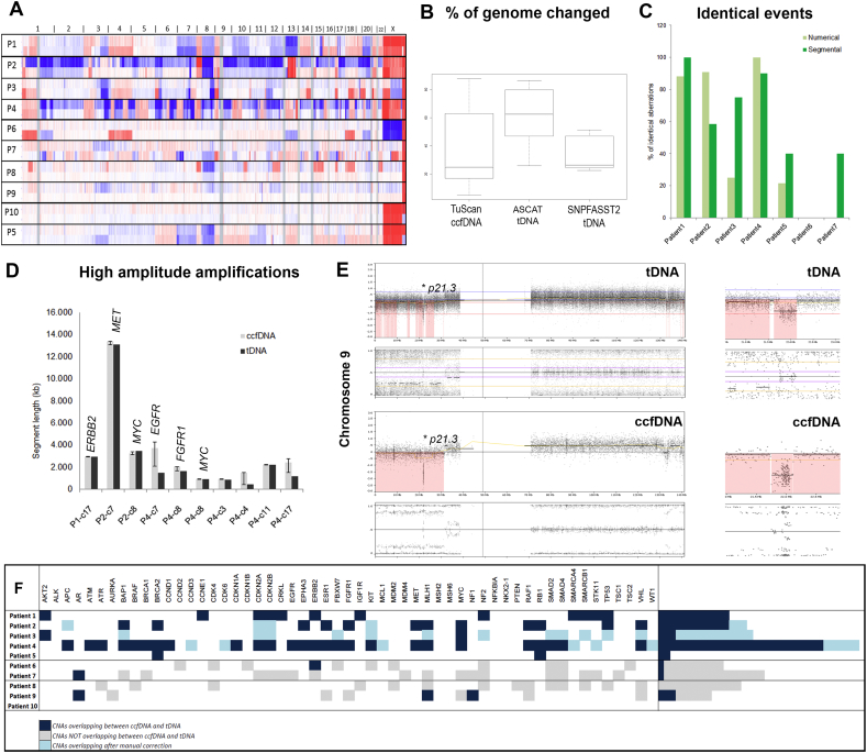
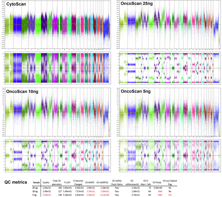
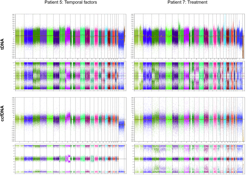
Methods: SNP microarrays (Affymetrix) were used to generate whole-genome copy number profiles from plasma ccfDNA (OncoScan) and paired tumor biopsies (CytoScan) from ten patients with metastatic cancers. Numerical, segmental and focal SCNAs were assessed using ASCAT/TuScan and SNP-FASST2.
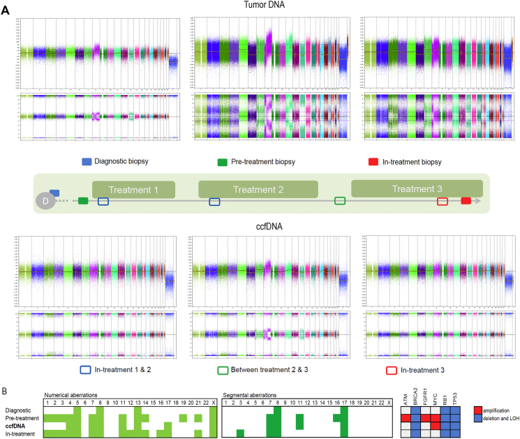
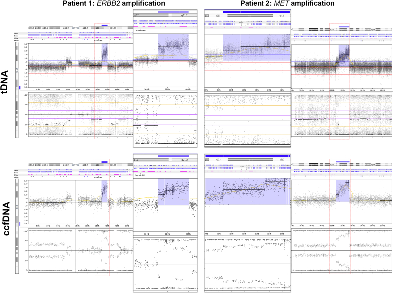
Results: Aberrations in ccfDNA in 4 patients resembled numerical (76%) and segmental (80%) aberrations in tDNA. Three patients represented low correlation due to postponed sampling time, ccfDNA quality and possible treatment interference. Breakpoints of high-amplitude amplification were assessed with high accuracy and relative breakpoints difference of only 7% (0.02-37%). Similarly, biallelic losses were reliably detected. Array was 100% successful in detection of SCNAs on clinically relevant genes compared to SCNAs in tumor biopsies. Tracking of SCNAs changes during the treatment course of one patient also indicated that apoptosis/necrosis of non-cancerous cells presumably induced by treatment can influence ccfDNA composition and introduce false-negative findings into the analysis of liquid biopsies.
Conclusions: Genomic alterations detected in ccfDNA from liquid biopsies by comprehensive SNP array are reliable source for information for stratification of patients for targeted treatment.
General significance: Clinically relevant SCNAs can be detected in ccfDNA with high resolution and can therefore serve as an alternative to tumor biopsy in defining treatment targets.
Keywords: Array profiling; Circulating cell-free tumor DNA; Copy number alterations; Diagnostics.
Figures
References
-
- Bettegowda C., Sausen M., Leary R.J., Kinde I., Wang Y., Agrawal N., Bartlett B.R., Wang H., Luber B., Alani R.M., Antonarakis E.S., Azad N.S., Bardelli A., Brem H., Cameron J.L., Lee C.C., Fecher L.A., Gallia G.L., Gibbs P., Le D., Giuntoli R.L., Goggins M., Hogarty M.D., Holdhoff M., Hong S.M., Jiao Y., Juhl H.H., Kim J.J., Siravegna G., Laheru D.A., Lauricella C., Lim M., Lipson E.J., Marie S.K., Netto G.J., Oliner K.S., Olivi A., Olsson L., Riggins G.J., Sartore-Bianchi A., Schmidt K., Shih I.M., Oba-Shinjo S.M., Siena S., Theodorescu D., Tie J., Harkins T.T., Veronese S., Wang T.L., Weingart J.D., Wolfgang C.L., Wood L.D., Xing D., Hruban R.H., Wu J., Allen P.J., Schmidt C.M., Choti M.A., Velculescu V.E., Kinzler K.W., Vogelstein B., Papadopoulos N., Diaz L.A., Jr. Detection of circulating tumor DNA in early- and late-stage human malignancies. Sci. Transl. Med. 2014;6 - PMC - PubMed
-
- Bolli N., Avet-Loiseau H., Wedge D.C., Van Loo P., Alexandrov L.B., Martincorena I., Dawson K.J., Iorio F., Nik-Zainal S., Bignell G.R., Hinton J.W., Li Y., Tubio J.M., McLaren S., S O.M., Butler A.P., Teague J.W., Mudie L., Anderson E., Rashid N., Tai Y.T., Shammas M.A., Sperling A.S., Fulciniti M., Richardson P.G., Parmigiani G., Magrangeas F., Minvielle S., Moreau P., Attal M., Facon T., Futreal P.A., Anderson K.C., Campbell P.J., Munshi N.C. Heterogeneity of genomic evolution and mutational profiles in multiple myeloma. Nat. Commun. 2014;5:2997. - PMC - PubMed
-
- Bronkhorst A.J., Aucamp J., Pretorius P.J. Cell-free DNA: preanalytical variables. Clin. Chim. Acta. 2015;450:243–253. - PubMed
-
- Chan K.C., Jiang P., Zheng Y.W., Liao G.J., Sun H., Wong J., Siu S.S., Chan W.C., Chan S.L., Chan A.T., Lai P.B., Chiu R.W., Lo Y.M. Cancer genome scanning in plasma: detection of tumor-associated copy number aberrations, single-nucleotide variants, and tumoral heterogeneity by massively parallel sequencing. Clin. Chem. 2013;59:211–224. - PubMed
-
- Chicard M., Boyault S., Colmet Daage L., Richer W., Gentien D., Pierron G., Lapouble E., Bellini A., Clement N., Iacono I., Brejon S., Carrere M., Reyes C., Hocking T., Bernard V., Peuchmaur M., Corradini N., Faure-Conter C., Coze C., Plantaz D., Defachelles A.S., Thebaud E., Gambart M., Millot F., Valteau-Couanet D., Michon J., Puisieux A., Delattre O., Combaret V., Schleiermacher G. Genomic copy number profiling using circulating free tumor DNA highlights heterogeneity in neuroblastoma. Clin. Cancer Res. 2016;22:5564–5573. - PubMed
LinkOut - more resources
Full Text Sources
Other Literature Sources
Molecular Biology Databases
