

Potential Coagulation Factor-Driven Pro-Inflammatory Responses in Ovarian Cancer Tissues Associated with Insufficient O₂ and Plasma Supply
- PMID: 28417928
- PMCID: PMC5412393
- DOI: 10.3390/ijms18040809
Potential Coagulation Factor-Driven Pro-Inflammatory Responses in Ovarian Cancer Tissues Associated with Insufficient O₂ and Plasma Supply
Abstract
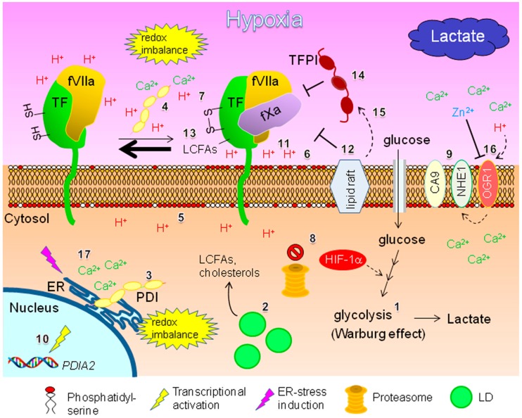
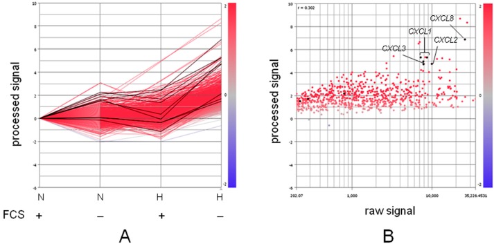
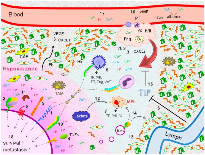
Tissue factor (TF) is a cell surface receptor for coagulation factor VII (fVII). The TF-activated fVII (fVIIa) complex is an essential initiator of the extrinsic blood coagulation process. Interactions between cancer cells and immune cells via coagulation factors and adhesion molecules can promote progression of cancer, including epithelial ovarian cancer (EOC). This process is not necessarily advantageous, as tumor tissues generally undergo hypoxia due to aberrant vasculature, followed by reduced access to plasma components such as coagulation factors. However, hypoxia can activate TF expression. Expression of fVII, intercellular adhesion molecule-1 (ICAM-1), and multiple pro-inflammatory cytokines can be synergistically induced in EOC cells in response to hypoxia along with serum deprivation. Thus, pro-inflammatory responses associated with the TF-fVIIa-ICAM-1 interaction are expected within hypoxic tissues. Tumor tissue consists of multiple components such as stromal cells, interstitial fluid, albumin, and other micro-factors such as proton and metal ions. These factors, together with metabolism reprogramming in response to hypoxia and followed by functional modification of TF, may contribute to coagulation factor-driven inflammatory responses in EOC tissues. The aim of this review was to describe potential coagulation factor-driven inflammatory responses in hypoxic EOC tissues. Arguments were extended to clinical issues targeting this characteristic tumor environment.
Keywords: coagulation; hypoxia; inflammation; ovarian cancer.
Conflict of interest statement
The authors declare no conflicts of interest.
Figures
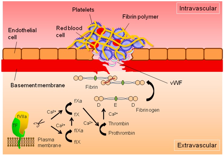
 : cleavage;
  : conversion.
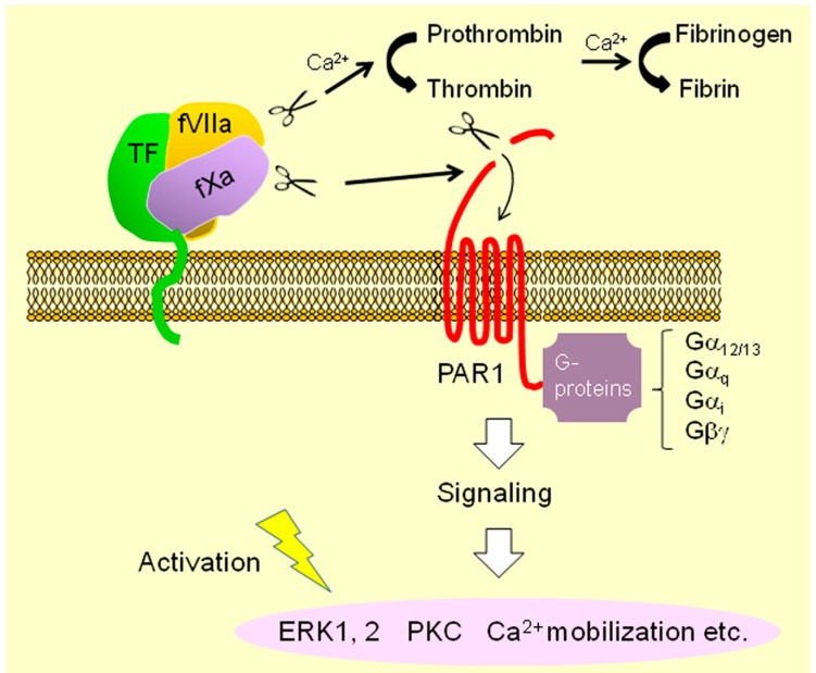 : cleavage; : conversion;
  : binding;
  : intracellular signaling.
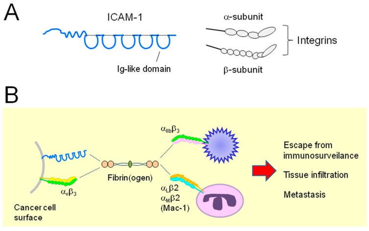
 : secretion;
  : interaction;
  : intracellular signaling.
References
-
- Ovarian Cancer National Alliance Statistics (webpage on the Internet) [(accessed on 31 January 2017)];2017 Washington, DC: Ovarian Cancer Research Fund Alliance. Available online: https://ocrfa.org.
-
- Anglesio M.S., Wiegand K.C., Melnyk N., Chow C., Salamanca C., Prentice L.M., Senz J., Yang W., Spillman M.A., Cochrane D.R. Type-specific cell line models for type-specific ovarian cancer research. PLoS ONE. 2013;8:e72162. doi: 10.1371/annotation/856f0890-9d85-4719-8e54-c27530ac94f4. - DOI - PMC - PubMed
-
- Chang C.-M., Chuang C.-M., Wang M.-L., Yang Y.-P., Chuang J.-H., Yang M.-J., Yen M.-S., Chiou S.-H., Chang C.-C. Gene set—Based integrative analysis revealing two distinct functional regulation patterns in four common subtypes of epithelial ovarian cancer. Int. J. Mol. Sci. 2016;17:1272. doi: 10.3390/ijms17081272. - DOI - PMC - PubMed
Publication types
MeSH terms
Substances
LinkOut - more resources
Full Text Sources
Other Literature Sources
Medical
Miscellaneous
