. 2017 Jul;108(7):1421-1431.
doi: 10.1111/cas.13260. Epub 2017 May 31.

Endoplasmic reticulum stress pathway PERK-eIF2α confers radioresistance in oropharyngeal carcinoma by activating NF-κB

Affiliations

Endoplasmic reticulum stress pathway PERK-eIF2α confers radioresistance in oropharyngeal carcinoma by activating NF-κB

Qiao Qiao et al. Cancer Sci. 2017 Jul.

Abstract

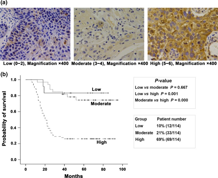
Endoplasmic reticulum stress (ERS) plays an important role in the pathogenesis and development of malignant tumors, as well as in the regulation of radiochemoresistance and chemoresistance in many malignancies. ERS signaling pathway protein kinase RNA-like endoplasmic reticulum kinase (PERK)-eukaryotic initiation factor-2 (eIF2α) may induce aberrant activation of nuclear factor-κB (NF-κB). Our previous study showed that NF-κB conferred radioresistance in lymphoma cells. However, whether PERK-eIF2α regulates radioresistance in oropharyngeal carcinoma through NF-κB activation is unknown. Herein, we showed that PERK overexpression correlated with a poor prognosis for patients with oropharyngeal carcinoma (P < 0.01). Meanwhile, the percentage of the high expression level of PERK in oropharyngeal carcinoma patients resistant to radiation was higher than in patients sensitive to radiation (77.7 and 33.3%, respectively; P < 0.05). Silencing PERK and eIF2α increased the radiosensitivity in oropharyngeal carcinoma cells and increased radiation-induced apoptosis and G2/M phase arrest. PERK-eIF2α silencing also inhibited radiation-induced NF-κB phosphorylation and increased the DNA double strand break-related proteins ATM phosphorylation. NF-κB activator TNF-α and the ATM inhibitor Ku55933 offset the regulatory effect of eIF2α on the expression of radiation-induced cell apoptosis-related proteins and the G2/M phase arrest-related proteins. These data indicate that PERK regulates radioresistance in oropharyngeal carcinoma through NF-kB activation-mediated phosphorylation of eIF2α, enhancing X-ray-induced activation of DNA DSB repair, cell apoptosis inhibition and G2/M cell cycle arrest.

Keywords: PERK; NF-κB; eIF2α; oropharyngeal carcinoma; radiotherapy.


Figures

Figure 1
Figure 1
Expression of PERK protein in human oropharyngeal carcinoma samples. (a) Schematic diagram of high, moderate and low expression of PERK protein in the cytoplasm of oropharyngeal carcinoma samples. (b) The Kaplan–Meier analysis showed that overall survival of patients with oropharyngeal carcinoma had a significant correlation with PERK protein expression level.
Figure 2
Figure 2
Expression of PERKeIF2α in radioresistant oropharyngeal carcinoma cells. (a) Colony formation results showing that the colony formation rates of FaDuR and Detroit562R were higher than those of FaDuP and Detroit562P after irradiation. (b) Western blot results showing that the expression levels of PERK and phospho‐eIF2α in radioresistant cells, FaDuR and Detroit562R were higher than those in FaDuP and Detroit562P. The bands were quantified using ImageJ software and normalized against a loading control. Changes in expression are shown in comparison to FaDuP and Detroit562P cells. NA, not applicable.
Figure 3
Figure 3
Expression of PERKeIF2α in oropharyngeal carcinoma cells after irradiation. (a) Western blot results showing that the expression of PERKeIF2α was activated in oropharyngeal carcinoma cells at different time points after 5 Gy irradiation. Bands were quantified using ImageJ software and normalized to a loading control. Fold changes are shown in comparison to the control lane. (b) Real‐time quantitative PCR results showing that the expression of PERKeIFmRNA increased 1 h after 5 Gy irradiation. Compared with the control group, *P < 0.05, #P < 0.01. NA, not applicable.
Figure 4
Figure 4
Effects of PERK on radiosensitivity in human oropharyngeal carcinoma cells. (a) Western blot results showing that transfection of PERK siRNA effectively inhibited PERK protein expression and radiation‐induced PERK protein expression. (b) Colony formation results showing that PERK silencing reduced the colony formation rates after radiotherapy. (c) Western blot results showing that 12 h after 5 Gy irradiation, PERK silencing inhibited eIF2α phosphorylation and Bcl‐2 protein expression, and increased ATM phosphorylation and cleaved caspase‐3 protein expression. The bands were quantified using ImageJ software and normalized to a loading control. Fold changes are shown relative to the negative control lane without radiation. NA, not applicable.
Figure 5
Figure 5
Effects of eIF2α phosphorylation on radiosensitivity in oropharyngeal carcinoma cells. (a) Western blot results showing that transfection of eIF2α siRNA effectively inhibited the cellular eIF2α phosphorylation level and radiation‐induced eIF2α phosphorylation. (b) Colony formation results showing that eIF2α silencing reduced colony formation rates after radiotherapy. (c) Western blot results showing that silencing of IRE and ATF‐6 did not affect eIF2α phosphorylation. Bands were quantified using ImageJ software and normalized to a loading control. Fold changes are shown in comparison to the negative control lane without radiation. NA, not applicable.
Figure 6
Figure 6
eIF2α regulated radioresistance in oropharyngeal carcinoma cells by activating the NF‐κB pathway. Cells were transfected with siRNA to silence eIF2α and irradiated with 5 Gy X‐ray. (a) Cells were collected after 24 h to detect apoptosis using Annexin V/PI staining followed by flow cytometry analysis. (b) Cells were collected after 48 h to identify the cell cycle using PI staining followed by flow cytometry. The results are expressed as the mean ± SD of three independent experiments. Compared with the IR group, *P < 0.05, #P < 0.01. (c) Cells were transfected with siRNA to silence eIF2α and irradiated with 5 Gy for 12 h. Nuclear and cytoplasm proteins were extracted to detect the expression of phospho‐p65 and its downstream protein HIF‐1α. Total protein was extracted to evaluate the expression of Rad50, Mre11, Nbs1 and phospho‐ATM proteins. Western blot results showed that eIF2α silencing inhibited radiation‐induced nuclear phospho‐p65 protein expression. Conversely, radiation inhibited cytoplasmic p65 protein phosphorylation, whereas eIF2α silencing reversed this function. These results indicated that eIF2α silencing inhibited radiation‐induced p65 nuclear translocation. In addition, eIF2α silencing inhibited the radiation‐induced HIF‐1α, Rad50 and Nbs1 expression, increased radiation‐induced phospho‐ATM expression, and did not affect Mre11 protein expression. (d) After eIF2α was silenced by siRNA transfection, the cells were irradiated (5 Gy, 1 h). The real‐time quantitative PCR results showed that eIF2α silencing decreased the radiation‐induced expression of p65 mRNA expression. Compared to the irradiated group, *P < 0.05 and # P < 0.01. (e) Immunofluorescence studies showed that after oropharyngeal carcinoma cells received 5 Gy radiation for 1 h, the γ‐H2AX foci in nucleus increased (the blue background indicates the cell nucleus, and light red dots indicate γ‐H2AX foci). In addition, the effect of radiation after eIF2α silencing was more evident than that of simple radiation. (f) The western blot results showed that eIF2α silencing inhibited radiation (5 Gy, 12 h)‐induced phospho‐cdc2, Cyclin B, Bcl‐2 and Bcl‐xL protein expression and increased radiation‐induced cleaved‐Caspase 3 and cleaved‐PARP protein expression. The above functions were inhibited by pretreatment of the oropharyngeal carcinoma cells with the NF‐κB activator TNFα (10 ng/mL) and the ATM inhibitor Ku55933 (10 μmol/L) for 12 h. Bands were quantified using ImageJ software and normalized to a loading control. Fold changes are shown compared with the negative control lane without radiation. NA, not applicable.

References

    1. Jemal A, Siegel R, Ward E et al Cancer statistics, 2008. CA Cancer J Clin 2008; 58: 71–96. - PubMed
    1. Harding HP, Zhang Y, Bertolotti A, Zeng H, Ron D. Perk is essential for translational regulation and cell survival during the unfolded protein response. Mol Cell 2000; 5: 897–904. - PubMed
    1. Ma Y, Hendershot LM. The role of the unfolded protein response in tumour development: friend or foe? Nat Rev Cancer 2004; 4: 966–77. - PubMed
    1. Qi L, Wu P, Zhang X et al Inhibiting ERp29 expression enhances radiosensitivity in human nasopharyngeal carcinoma cell lines. Med Oncol 2012; 29: 721–8. - PubMed
    1. Nagelkerke A, Bussink J, van der Kogel AJ, Sweep FC, Span PN. The PERK/ATF4/LAMP3‐arm of the unfolded protein response affects radioresistance by interfering with the DNA damage response. Radiother Oncol 2013; 108: 415–21. - PubMed

MeSH terms