Review
. 2017 May;6(5):293-304.
doi: 10.1002/psp4.12191. Epub 2017 Apr 18.

Towards Quantitative Systems Pharmacology Models of Chemotherapy-Induced Neutropenia

Affiliations
Review

Towards Quantitative Systems Pharmacology Models of Chemotherapy-Induced Neutropenia

M Craig. CPT Pharmacometrics Syst Pharmacol. 2017 May.

Abstract

Neutropenia is a serious toxic complication of chemotherapeutic treatment. For years, mathematical models have been developed to better predict hematological outcomes during chemotherapy in both the traditional pharmaceutical sciences and mathematical biology disciplines. An increasing number of quantitative systems pharmacology (QSP) models that combine systems approaches, physiology, and pharmacokinetics/pharmacodynamics have been successfully developed. Here, I detail the shift towards QSP efforts, emphasizing the importance of incorporating systems-level physiological considerations in pharmacometrics.


Figures

Figure 1
Figure 1
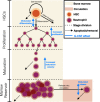
An overview of granulopoiesis. As with all blood cells, neutrophils begin as hematopoietic stem cells (HSCs, orange circle) in the bone marrow (pale yellow background), where they develop. HSCs are capable of self‐renewal and are subject to cell death (dashed arrows). HSCs may also differentiate into one of the blood cell lines, including the neutrophils (purple circles). After commitment to the neutrophil lineage, cells undergo a period of proliferative expansion at the end of which they no longer divide. Postmitotic neutrophils then mature, growing in size and gaining receptors. At the end of the maturation process, cells are then stored in the bone marrow reservoir from which they egress to reach the circulation (pale red background) before removal (by margination or death). G‐CSF acts to modulate the rate of exit from the marrow reservoir, increase the rates of maturation and proliferation, and to modulate the rate of differentiation into the neutrophil lineage (G‐CSF actions represented by blue vertical arrows).
Figure 2
Figure 2
Schematic representation of the general semimechanistic model of neutrophil development developed by Friberg.39 Proliferative cells self‐renew at rate kprol or begin the transition to the circulation by exiting with rate ktr (the rate of transit). The delay between the time cells leave proliferation to when they enter the circulation is called the mean transit time and is equally divided between n transit compartment, each connected by the rate of transit. The original model as presented in Friberg39 and used most frequently is restricted to three transit compartments. More recent extensions have more recently appeared.40, 41 Once cells enter the circulation, they are removed with rate kcirc. The number of circulating cells has a negative feedback on the proliferation rate of the proliferative cells. The myelosuppressive action of the drug is assumed to also affect kprol.
Figure 3
Figure 3
The integrated G‐CSF‐myelosuppression model describing the dynamics of endogenous G‐CSF and neutrophils following chemotherapy. For the myelosuppression model the parameters are baseline neutrophil count (ANC0), mean maturation time (MMT = 5/ktr), the half‐life of neutrophils in circulation (t1/2circ = LN(2)/kcirc), the feedback parameters of G‐CSF on neutrophil proliferation (γ) and transit time (β) and the drug‐related effect (Edrug). The estimated parameters for the G‐CSF turnover model are baseline G‐CSF (GCSF0), nonspecific elimination rate constant (ke), and ANC‐dependent elimination rate constant (kANC) and cortisol‐induced G‐CSF release (DOSEcort) and the half‐life of cortisol‐induced G‐CSF release (t1/2cort = LN(2)/kcort). Figure reproduced from “Characterization of endogenous G‐CSF and the inverse correlation to chemotherapy‐induced neutropenia in patients with breast cancer using population modeling,” Pharm. Res. 31, 2014, p. 3396, Quartino, A.L. et al.,41 with the permission of Springer.
Figure 4
Figure 4
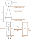
Schematic representation of the production of circulating neutrophils in the bone marrow and the interaction of the system with G‐CSF. Hematopoietic stem cells (HSCs‐Q) enter the neutrophil lineage, the other blood lines, or are removed from the HSC pool. Differentiated HSCs undergo successive divisions during the proliferative phase. Cells then mature before being stored in the marrow reservoir, or dying off during maturation. Neutrophils remain in the reservoir until they are removed randomly or enter the circulation, where they disappear rapidly from the blood. Freely circulating G‐CSF may bind to receptors on the neutrophils. The concentration of bound G‐CSF drives its pharmacodynamic effects. The concentration of G‐CSF bound to mature neutrophils, G 2, determines the rate of release from the marrow reservoir. The concentration of G‐CSF bound to neutrophil precursors, assumed proportional to G 1, the concentration of freely circulating G‐CSF, determines the rate of differentiation from the HSCs, the speed of maturation, and the rate of proliferation. For all four effects, speed and rates increase with increasing G‐CSF concentration. Figure reproduced from “A mathematical model of granulopoiesis incorporating the negative feedback dynamics and kinetics of G‐CSF/neutrophil binding and internalization,” Bull. Math. Biol., 78, 2016, p. 2308, Craig, M., Humphries, A.R., and Mackey, M.C.30 with the permission of Springer.

References

    1. Mantovani, A. , Cassatella, M.A. , Costantini, C. & Jaillon, S. Neutrophils in the activation and regulation of innate and adaptive immunity. Nat. Rev. Immunol. 11, 519–531 (2011). - PubMed
    1. von Vietinghoff, S. & Ley, K. Homeostatic regulation of blood neutrophil counts. J. Immunol. 181(8), 5183–5188 (2008). - PMC - PubMed
    1. Pillay, J. et al In vivo labeling with 2H2O reveals a human neutrophil lifespan of 5.4 days. Blood 116(4), 625–627 (2010). - PubMed
    1. Craig, M. , Humphries, A.R. & Mackey, M.C. An upper bound for the half‐removal time of neutrophils from circulation. Blood 128(15), 1989–1991 (2016). - PMC - PubMed
    1. Lahoz‐Beneytez, J. et al Human neutrophil kinetics: modeling of stable isotope labeling data supports short blood neutrophil half‐lives. Blood 127(26), 3431–3438 (2016). - PMC - PubMed

Publication types

Substances

LinkOut - more resources