

Methods for the isolation and 3D culture of dermal papilla cells from human hair follicles
- PMID: 28418608
- PMCID: PMC5519926
- DOI: 10.1111/exd.13368
Methods for the isolation and 3D culture of dermal papilla cells from human hair follicles
Abstract
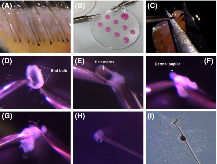
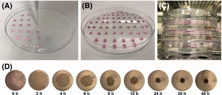
The dermal papilla is a cluster of mesenchymal cells located at the base of the hair follicle which have a number of important roles in the regulation of hair growth. As a consequence, in vitro models of these cells are widely used to study the molecular mechanisms which underlie hair follicle induction, growth and maintenance. While dermal papilla from rodent hair follicles can be digested prior to cell isolation, the unique extracellular matrix composition found in human dermal papilla renders enzymes such as trypsin and collagenase insufficient for digestion of the dermal papilla into a single cell suspension. As such, to grow human dermal papilla cells in vitro, the papilla has to first be isolated via a micro-dissection approach from the follicle. In this article we describe the micro-dissection and culture methods, which we use within our laboratory, for the study of human dermal papilla cells.
Keywords: dermal papilla; hair follicle; inversion; micro-dissection; spheroid culture.
© 2017 The Authors. Experimental Dermatology Published by John Wiley & Sons Ltd.
Figures
References
Publication types
MeSH terms
Grants and funding
LinkOut - more resources
Full Text Sources
Other Literature Sources
Research Materials
