Review
. 2017 Jul 25:40:211-230.
doi: 10.1146/annurev-neuro-072116-031335. Epub 2017 Apr 18.

Visual Circuits for Direction Selectivity

Affiliations
Review

Visual Circuits for Direction Selectivity

Alex S Mauss et al. Annu Rev Neurosci. .

Abstract

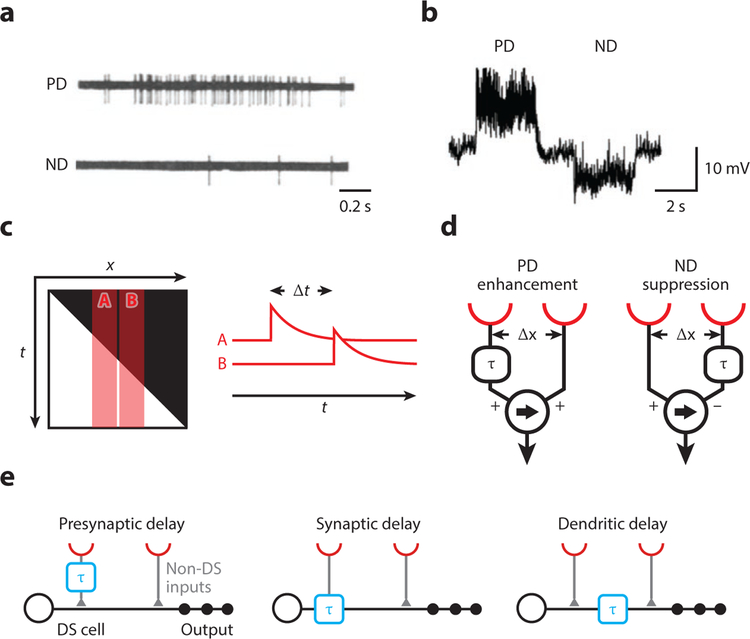
Images projected onto the retina of an animal eye are rarely still. Instead, they usually contain motion signals originating either from moving objects or from retinal slip caused by self-motion. Accordingly, motion signals tell the animal in which direction a predator, prey, or the animal itself is moving. At the neural level, visual motion detection has been proposed to extract directional information by a delay-and-compare mechanism, representing a classic example of neural computation. Neurons responding selectively to motion in one but not in the other direction have been identified in many systems, most prominently in the mammalian retina and the fly optic lobe. Technological advances have now allowed researchers to characterize these neurons' upstream circuits in exquisite detail. Focusing on these upstream circuits, we review and compare recent progress in understanding the mechanisms that generate direction selectivity in the early visual system of mammals and flies.

Keywords: direction selectivity; motion detection; optic lobe; retina; visual system.


Figures

Figure 1
Figure 1. Cells and models for visual motion detection.
(a) Spiking response of a direction-selective unit in the rabbit retina to a moving light spot. Panel adapted with permission from Barlow and Levick (1965). (b) Response of a lobula plate tangential cell in Drosophila to a grating moving in opposite directions. Panel adapted with permission from Joesch et al. (2008). (c) The brightness profile of a light edge moving to the right shown in space-time (x-t). A and B represent the receptive fields of two adjacent photoreceptors, which are activated sequentially with a delay Δt. The traces A and B depict a high-pass-filtered version of a signal, highlighting illumination changes. (d) Two alternative motion detector models. Both generate direction-selective responses by a delay-and-compare mechanism but differ in their nonlinear integration. In the Hassenstein-Reichardt model on the left, a delayed signal (denoted by τ) enhances a direct signal, for instance by a multiplication. In the Barlow-Levick model on the right, a delayed signal suppresses a direct signal, for instance by a division. In both models, the arrow indicates the preferred direction. (e) Potential cellular implementations of local motion detection (not mutually exclusive). Spatially offset signals are conveyed through different cell types or different synaptic receptors with different dynamics (left and middle, respectively). Temporal delays might also arise by dendritic filtering (right). Note that “presynaptic delay” could be generated by an arbitrary mechanism in the upstream circuit. Abbreviations: DS, direction-selective; ND, null-direction; PD, preferred-direction.
Figure 2
Figure 2. Visual motion detection in the mammalian retina.
(a) Schematic of the mammalian retinal circuit elements for direction selectivity. Photoreceptor signals are split into an ON and an OFF pathway at the level of bipolar cells via different glutamate receptors. ON and OFF bipolar cells synapse individually onto direction-selective ON and OFF SACs, respectively, and jointly onto ON-OFF DSGCs. DSGC receive additional asymmetric GABAergic and symmetric cholinergic input from SACs. Note that only selected aspects of the connectivity are captured in this simplified schematic. (b) A z-projection of an ON SAC filled with a fluorescent dye showing the radial dendrites and stratification. (c) Distribution of inputs and outputs determined by serial electron microscopy reconstructions of mouse ON SACs as well as their pre- and postsynaptic partners. Panel adapted with permission from Ding et al. (2016). (d) (left) Distributions of glutamatergic inputs on SAC dendrites determined using glutamate uncaging. Each row is a different SAC dendrite, the solid line is the mapped region of the dendrite, and blue dots are glutamatergic synapses as revealed by glutamate uncaging. (right) Comparison of glutamatergic inputs determined by labeling SACs with the postsynaptic marker for glutamatergic synapses, PSD95-YFP (blue) and outputs determined using serial electron microscopy reconstructions. The average location of the most distal synapse measured with uncaging is represented by the dotted line. Panel adapted with permission from Vlasits et al. (2016). (e) Schematic of dendritic locations of inputs (blue, bipolar cells; red, neighboring SACs) and outputs (black) in mouse and rabbit SACs. Abbreviations: AC, amacrine cell; BC, bipolar cell; DSGC, direction-selective ganglion cell; EM, electron microscopy; PR, photoreceptor; SAC, starburst amacrine cell.
Figure 3
Figure 3. Visual motion detection in the Drosophila optic lobe.
(a) Schematic representation of the motion detection circuit. Photoreceptor signals are split into an ON (left) and an OFF (right) pathway processing motion information independently. Lamina (L) and medulla (Mi and Tm) cells constitute different temporal filters whose spatially offset outputs are integrated in the T4 and T5 dendrites. There, direction-selective signals are generated. T4/T5 cells thus constitute local motion detectors. Note that the spatial arrangement of T4/T5 inputs as shown here is meaningless and that inputs to leftward-tuned T4/T5 cells are omitted for clarity. Tangential cells of the lobula plate integrate direct excitatory T4/T5 signals and indirect sign-inverted signals via LPi neurons with opposite tuning (illustrated by opposing arrows) across large receptive fields. (b) Dendrite of a T4 cell (green) tuned to upward motion. Medulla columns representing individual points in visual space are labeled with a synaptic marker in magenta. Note that the T4 dendrite spreads across multiple columns. Image courtesy of J. Pujol-Marti. (c) Image of compound eye illuminated from the inside. This can be implemented to precisely stimulate single points in fly visual space, individually or sequentially. (d) Calcium signals of a T4 cell, obtained by two-photon imaging. Sequential stimulation (apparent motion) of three adjacent pixels in an upward direction (A to C) produces a combined response, which is larger than the sum of the three individual responses (superlinear). Conversely, apparent downward motion (C to A) produces a combined response, which is smaller than the sum of the three individual responses (sublinear). (e) T4 cells generate direction-selective signals by two complementary mechanisms: preferred-direction excitation and null-direction suppression. (f) In simulations, null-direction suppression or preferred-direction enhancement alone produce broadly tuned direction-selective responses. A detector combining both elements yields markedly increased direction selectivity (matching experimentally obtained data), highlighting a potential benefit for combining two mechanisms. Panels c–f adapted with permission from Haag et al. (2016). (g) Responses to brightness decrements of varying durations of the four nondirection-selective inputs to direction-selective T5 cells, measured by two-photon calcium imaging. Tm cells can be characterized as fast and transient (Tm2 and Tm4), intermediate (Tm1), and slow (Tm9). Onsets and offsets of light stimuli are indicated below; 0 represents light off, 1 represents light on. Panel adapted with permission from Serbe et al. (2016). Abbreviations: DF/F, delta fluorescence/fluorescence (relative change of calcium indicator fluorescence); L, lamina cell; LMD, local motion detector; LPi, lobula plate-intrinsic cell; Mi, medulla-intrinsic cell; PR, photoreceptor; TC, tangential cell; TF, temporal filter; Tm, transmedullary cell; WI, wide-field integrator.

Similar articles

Cited by

References

    1. Ammer G, Leonhardt A, Bahl A, Dickson BJ, Borst A. 2015. Functional specialization of neural input elements to the Drosophila ON motion detector. Curr. Biol 25(17):2247–53 - PubMed
    1. Amthor FR, Keyser KT, Dmitrieva NA. 2002. Effects of the destruction of starburst-cholinergic amacrine cells by the toxin AF64A on rabbit retinal directional selectivity. Vis. Neurosci 19(4):495–509 - PubMed
    1. Baden T, Berens P, Bethge M, Euler T. 2013. Spikes in mammalian bipolar cells support temporal layering of the inner retina. Curr. Biol 23(1):48–52 - PubMed
    1. Bahl A, Ammer G, Schilling T, Borst A. 2013. Object tracking in motion-blind flies. Nat. Neurosci 16(6):730–38 - PubMed
    1. Baines RA, Uhler JP, Thompson A, Sweeney ST, Bate M. 2001. Altered electrical properties in Drosophila neurons developing without synaptic transmission. J. Neurosci 21(5):1523–31 - PMC - PubMed