. 2017 Apr 18;13(4):e1006714.
doi: 10.1371/journal.pgen.1006714. eCollection 2017 Apr.

Microhomology-mediated end joining induces hypermutagenesis at breakpoint junctions

Affiliations

Microhomology-mediated end joining induces hypermutagenesis at breakpoint junctions

Supriya Sinha et al. PLoS Genet. .

Abstract

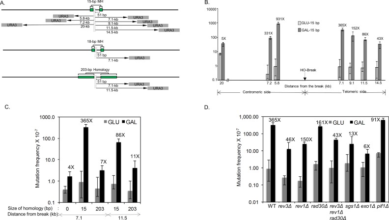
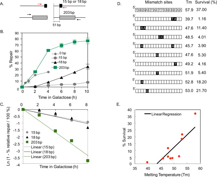
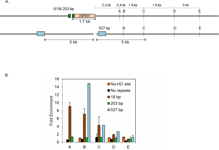
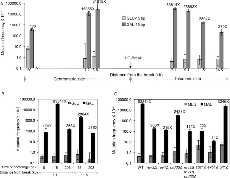
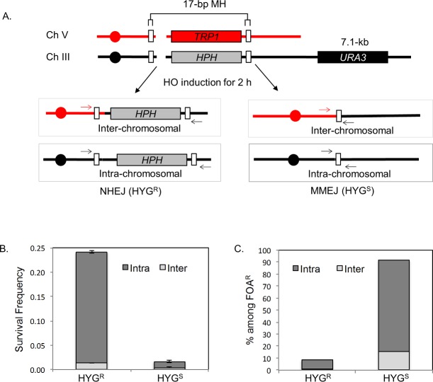
Microhomology (MH) flanking a DNA double-strand break (DSB) drives chromosomal rearrangements but its role in mutagenesis has not yet been analyzed. Here we determined the mutation frequency of a URA3 reporter gene placed at multiple locations distal to a DSB, which is flanked by different sizes (15-, 18-, or 203-bp) of direct repeat sequences for efficient repair in budding yeast. Induction of a DSB accumulates mutations in the reporter gene situated up to 14-kb distal to the 15-bp MH, but more modestly to those carrying 18- and 203-bp or no homology. Increased mutagenesis in MH-mediated end joining (MMEJ) appears coupled to its slower repair kinetics and the extensive resection occurring at flanking DNA. Chromosomal translocations via MMEJ also elevate mutagenesis of the flanking DNA sequences 7.1 kb distal to the breakpoint junction as compared to those without MH. The results suggest that MMEJ could destabilize genomes by triggering structural alterations and increasing mutation burden.


Conflict of interest statement

The authors have declared that no competing interests exist.

Figures

Fig 1
Fig 1. Microhomology-mediated repair induces hypermutagenesis.
A. MMEJ and SSA systems. The position of 15- or 18-bp MH and 203-bp repeats flanking HO recognition sequences are shown as green boxes. The grey boxes indicate URA3 reporter gene placed at several locations distal to an HO recognition site (5.8-, 7.1-, 7.2-, 9.1-, 11.5-, 14.5-, and 20-kb) from the break. The HML, HMR and URA3 genes are deleted to avoid gene conversion events. B. DNA break-induced mutation frequency was calculated by the median of the fluctuation tests using the number of FOAR survivors in yeast strains carrying the URA3 reporter gene placed at indicated locations distal to the HO break site. The HO recognition site, shown by the arrow, is flanked by 15-bp MH that mediates MMEJ repair upon induction of galactose inducible HO endonuclease. Distance from the break and the fold stimulation, calculated by dividing the mutation frequency of induced cells (gal; galactose) by that of uninduced (glu; glucose) controls are shown below and above the bar graph, respectively. Plotted in the graphs are the median frequencies, 95% confidence intervals, and fold change. The values are also listed in S2 Table. C. The frequency of FOAR survivors from yeast strains bearing 15-bp MH, 203-bp repeats or no homology flanking the HO break site, and the URA3 reporter gene placed at 7.1- and 11.5-kb distal locations. The median frequencies, 95% confidence intervals, and fold change are shown. D. The frequency of FOAR survivors in yeast strains with the indicated gene deletion was measured as described above. The median frequencies, 95% confidence intervals, and fold change are also listed in S2 Table.
Fig 2
Fig 2. Kinetics of MH-mediated repair.
A. Strategy to assess DSB repair kinetics. The black and grey boxes represent 15- or 18-bp microhomology (MH) and 203 bp homology flanking the HO-break site, respectively. The level of repair product was determined by quantitative real time PCR of genomic DNA isolated from an aliquot of cell culture after HO induction using primers flanking the repeats (red and black arrows). B. Graph showing the amount of repair products by annealing 203-bp homology or 0-, 15-, and 18-bp MH at indicated time points after HO-endonuclease induction. The results are the average of three independent experiments ± s.d. C. First order reaction kinetics of MMEJ products as a function of time post-HO expression. The slope represents the rate constant (k), which is constant regardless of MH sizes but is different in SSA between 203-bp repeats. D. Illustration of yeast strains with imperfect 18-bp MH. Black boxes indicate the position of base mismatches. The melting temperature (Tm) of each MH sequence and the percentage survival upon HO endonuclease induction are shown. Percent survival was calculated by dividing the number of colonies on YEP-galactose by the number of colonies on YEP-dextrose and multiplied by 100. E. Linear regression analysis of percentage survival vs. melting temperature (Tm) of strains carrying MH with one or more base mismatches. Percentage survival was positively correlated with Tm (p<0.05, R2 0.667445).
Fig 3
Fig 3. Extensive resection in MH-mediated repair.
A. Diagram demonstrating the strategy to measure the amount of DNA repair synthesis during MMEJ and SSA repair as a proxy for end resection. The HO-recognition site and the flanking MH or homologies (18-, 203- and 527-bp; black boxes) at various locations distal to the break trigger MMEJ or SSA. The locations of primers (A, B, C, D and E, 2.2-, 2.6-, 4.5-, 6.1- and 9.1-kb from HO break site) to detect BrdU incorporation by chromatin immunoprecipitation using anti-BrdU antibody in a strain with 527 bp repeat are shown. Strains with 0-, 18- and 203-bp MH or homologies have a 1.76-kb HPH gene incorporated 51 bp proximal to the HO recognition site. In these strains the total amount of resection is calculated by adding 1.76 kb (size of the HPH gene) to the distance of the primers from the HO recognition site. The distance between homologies and the HO recognition site and the extent of resection required to uncover homologies in strain with 527 bp homology are also included. B. Fold enrichment of BrdU incorporation at A, B, C and D locations in strains carrying no MH, 18-bp MH, 203-bp or 527-bp repeats was calculated by measuring the amount of BrdU incorporation after HO endonuclease induction divided by that under no-HO conditions as described in Materials and Methods. The results are the average of three independent experiments ± s.d.
Fig 4
Fig 4. UV-induced mutagenesis during MMEJ repair.
A. UV-induced mutation frequency was measured by scoring FOAR survivors in yeast strains carrying the URA3 reporter gene placed at indicated locations distal to the HO break site as described in Fig 1. The HO recognition site, shown by the arrow, is flanked by 15-bp MH that mediates MMEJ repair upon galactose induction of HO endonuclease. The distance from the break and the fold stimulation by DSB induction are shown below and above each bar graph, respectively. The median frequencies, 95% confidence intervals, and fold change are also listed in S6 Table. B. The frequency of UV induced FOAR survivors from yeast strains bearing 15-bp MH, 203-bp repeat or no homology flanking the HO break site and the URA3 reporter genes at placed at 7.1- and 11.5-kb distal locations. The median frequencies, 95% confidence intervals, and fold change are shown as in S6 Table. C. The frequency of UV-induced FOAR survivors was measured as described in Fig 1 in yeast strains with the indicated gene deletions and bearing 15-bp MH flanking the HO break and the URA3 reporter gene at the 7.1-kb distal location. The median frequencies, 95% confidence intervals, and fold change are also listed in S6 Table.
Fig 5
Fig 5. MH-induced mutagenesis at chromosomal translocation breakpoints.
A. Schematics illustrating the yeast strain that produces intra- or inter-chromosomal MMEJ or NHEJ upon HO expression. The strain has two HO recognition sites, one on Chromosome III and the other on Chromosome V. White boxes denote the location of 17-bp MH near the break site. HPH and TRP1 markers are shown. Four possible repair outcomes in this strain after DSB induction based on hygromycin sensitivity (HYGs) or resistance (HYGr) and the types of chromosomal joints are shown. The formation of chromosomal translocations was determined by PCR across the HO cleavage sites using primers annealed to two different chromosomes (arrows). B. Types of repair events among survivors. Survival frequency is calculated by dividing the number of survivors by the number of cells plated. A DSB was induced in the strain for 2 h by incubation in YEP-galactose and cells were plated on YEP-dextrose after serial dilution. The percentage of intra- and inter-chromosomal repair events was determined by PCR analysis of 100 colonies from each survivor. The results are the average of three independent experiments ± s.d. C. Types of FOAR survivors after HO expression. The percentage of intra- vs inter-chromosomal repair events and the status of the hygromycin resistance gene are plotted. A DSB was induced for 2 h and cells were plated onto YEP-dextrose and subsequently replica plated onto 5-Fluoroorotic Acid (5-FOA) plates. 100 colonies from each experiment were analyzed by PCR to detect intra-chromosomal or inter-chromosomal repair products. The results are the average of three independent experiments.

Similar articles

Cited by

References

    1. Sinha S, Villarreal D, Shim EY, Lee SE. Risky business: Microhomology-mediated end joining . Mutat Res. 2016. Epub 2016/01/23. - PMC - PubMed
    1. McVey M, Lee SE. MMEJ repair of double-strand breaks (director's cut): deleted sequences and alternative endings. Trends Genet. 2008;24(11):529–38. Epub 2008/09/24. doi: 10.1016/j.tig.2008.08.007 - DOI - PMC - PubMed
    1. Sfeir A, Symington LS. Microhomology-Mediated End Joining: A Back-up Survival Mechanism or Dedicated Pathway? Trends Biochem Sci. 2015;40(11):701–14. Epub 2015/10/07. PubMed Central PMCID: PMCPMC4638128. doi: 10.1016/j.tibs.2015.08.006 - DOI - PMC - PubMed
    1. Lieber MR, Lu H, Gu J, Schwarz K. Flexibility in the order of action and in the enzymology of the nuclease, polymerases, and ligase of vertebrate non-homologous DNA end joining: relevance to cancer, aging, and the immune system. Cell Res. 2008;18(1):125–33. Epub 2007/12/19. doi: 10.1038/cr.2007.108 - DOI - PubMed
    1. Lieber MR. Mechanisms of human lymphoid chromosomal translocations. Nat Rev Cancer. 2016;16(6):387–98. Epub 2016/05/26. doi: 10.1038/nrc.2016.40 - DOI - PMC - PubMed

MeSH terms

LinkOut - more resources