. 2017 Apr 18;12(4):e0174241.
doi: 10.1371/journal.pone.0174241. eCollection 2017.

Anti-proliferative effect of novel primary cetyl alcohol derived sophorolipids against human cervical cancer cells HeLa

Affiliations

Anti-proliferative effect of novel primary cetyl alcohol derived sophorolipids against human cervical cancer cells HeLa

Laxman Nawale et al. PLoS One. .

Abstract

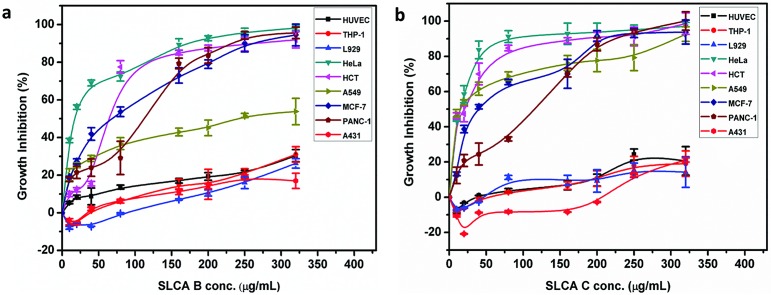
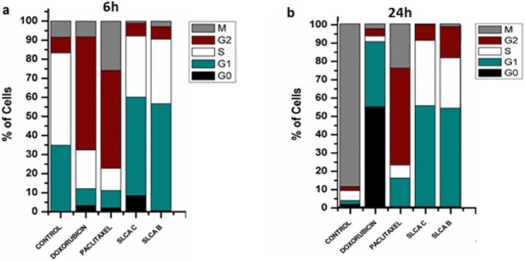
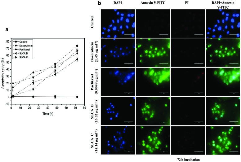
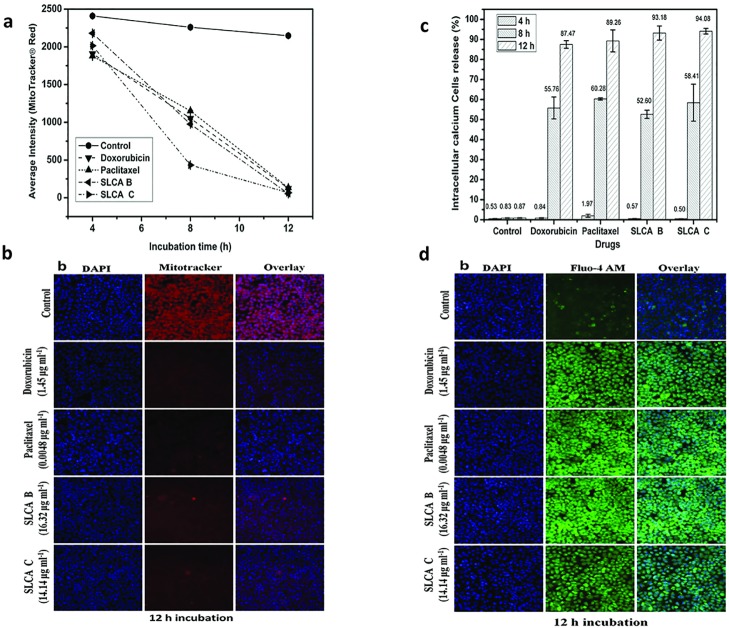
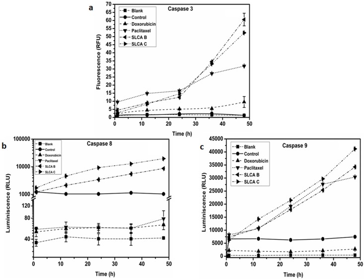
Sophorolipids (SLs) are glycolipid biosurfactants that have been shown to display anticancer activity. In the present study, we report anti-proliferative studies on purified forms of novel SLs synthesized using cetyl alcohol as the substrate (referred as SLCA) and their anticancer mechanism in human cervical cancer cells. Antiproliferative effect of column purified SLCA fractions (A, B, C, D, E and F) was examined in panel of human cancer cell lines as well as primary cells. Among these fractions, SLCA B and C significantly inhibited the survival of HeLa and HCT 116 cells without affecting the viability of normal human umbilical vein endothelial cells (HUVEC). The two fractions were identified as cetyl alcohol sophorolipids with non-hydroxylated tail differing in the degree of acetylation on sophorose head group. At an IC50 concentration SLCA B (16.32 μg ml-1) and SLCA C (14.14 μg ml-1) blocked the cell cycle progression of HeLa cells at G1/S phase in time-dependent manner. Moreover, SLCA B and SLCA C induced apoptosis in HeLa cells through an increase in intracellular Ca2+ leading to depolarization of mitochondrial membrane potential and increase in the caspase-3, -8 and -9 activity. All these findings suggest that these SLCAs could be explored for their chemopreventive potential in cervical cancer.


Conflict of interest statement

Competing Interests: The authors have declared that no competing interests exist.

Figures

Fig 1
Fig 1. SLCAs inhibits growth of cancer cells.
Cancer cell lines as well as primary HUVEC cells were subjected to different concentrations of SLCA B, SLCA C, doxorubicin and Paclitaxel for 48 h. Graphical representation of percent growth inhibition GI50 for (a) SLCA B (b) SLCA C. The data represents mean ± SD of three independent experiments.
Fig 2
Fig 2. SLCAs induce G1/S phase arrest in HeLa cells.
HeLa cells were subjected to different treatment at GI50 concentration for different time interval (6, 12, 18 and 14h). Post treatment, cells were monitored with DAPI staining and were analyzed by HCS. Graphical representation of % cells in different phases of cell cycle at 6h (a) and 24h (b).
Fig 3
Fig 3. SLCAs induce apoptosis in HeLa.
(a) Apoptotic ratio (b) Annexin-V (AV) and PI labeling of HeLa cells counterstained with DAPI. The overlay images in the second last and last column represent the apoptotic cells and absence of necrosis respectively. The analysis was conducted using confocal microscopy, Magnification 20X (scale, 100 μm).
Fig 4
Fig 4. SLCAs decrease mitochondrial membrane potential and increase intracellular calcium in HeLa cells.
Alteration of mitochondrial transmembrane potential in sophorolipid treated HeLa cells as measured by staining with Mito Tracker Red. (a) Average dye intensity signifying depolarized cells. Data from triplicate experiments represented as mean±SD (b) Fluorescence intensity of bound dye as recorded by confocal microscope (20X magnification, scale-100 μm). Elevation in intracellular [Ca2+]i on sophorolipid treatment of HeLa cells as measured by Fluo-4AM. (c) Bar diagram represents cell percentage releasing calcium at different time intervals. (d) Overlay of confocal microscopy images exemplifying increased Fluo-4 AM intensity after 12 h that indicates increase in cytoplasmic calcium. Magnification 20X (scale, 100 μm).
Fig 5
Fig 5. Effect of SLCA B and SLCA C on activation of caspases.
The sophorolipid treated cells were incubated for different time intervals and caspase activity was assessed by spectrometric method. (a) caspase-3, (b) caspase-8 and (c) caspase-9 activity was measured in comparison to the control. Data represented mean ± SD values.

References

    1. Van Bogaert INA, Saerens K, De Muynck C, Develter D, Soetaert W, Vandamme EJ. Microbial production and application of sophorolipids. Applied microbiology and biotechnology. 2007;76: 23–34. 10.1007/s00253-007-0988-7 - DOI - PubMed
    1. Chen J, Song X, Zhang H, Qu Y. Production, structure elucidation and anticancer properties of sophorolipid from Wickerhamiella domercqiae. Enzyme and Microbial Technology. 2006;39: 501–506.
    1. Shao L, Song X, Ma X-J, Li H, Qu Y. Bioactivities of sophorolipid with different structures against human esophageal cancer cells. The Journal of surgical research. 2012;173: 286–91. 10.1016/j.jss.2010.09.013 - DOI - PubMed
    1. Chen J, Song X, Zhang H, Qu YB, Miao JY. Sophorolipid produced from the new yeast strain Wickerhamiella domercqiae induces apoptosis in H7402 human liver cancer cells. Applied Microbiology and Biotechnology. 2006;72: 52–59. 10.1007/s00253-005-0243-z - DOI - PubMed
    1. Fu SL, Wallner SR, Bowne WB, Hagler MD, Zenilman ME, Gross RA, et al. Sophorolipids and their derivatives are lethal against human pancreatic cancer cells. The Journal of Surgical Research. 2008;148: 77–82. 10.1016/j.jss.2008.03.005 - DOI - PubMed

MeSH terms