. 2017 Jun 15;26(12):2207-2217.
doi: 10.1093/hmg/ddx110.

P4HA1 mutations cause a unique congenital disorder of connective tissue involving tendon, bone, muscle and the eye

Affiliations

P4HA1 mutations cause a unique congenital disorder of connective tissue involving tendon, bone, muscle and the eye

Yaqun Zou et al. Hum Mol Genet. .

Abstract

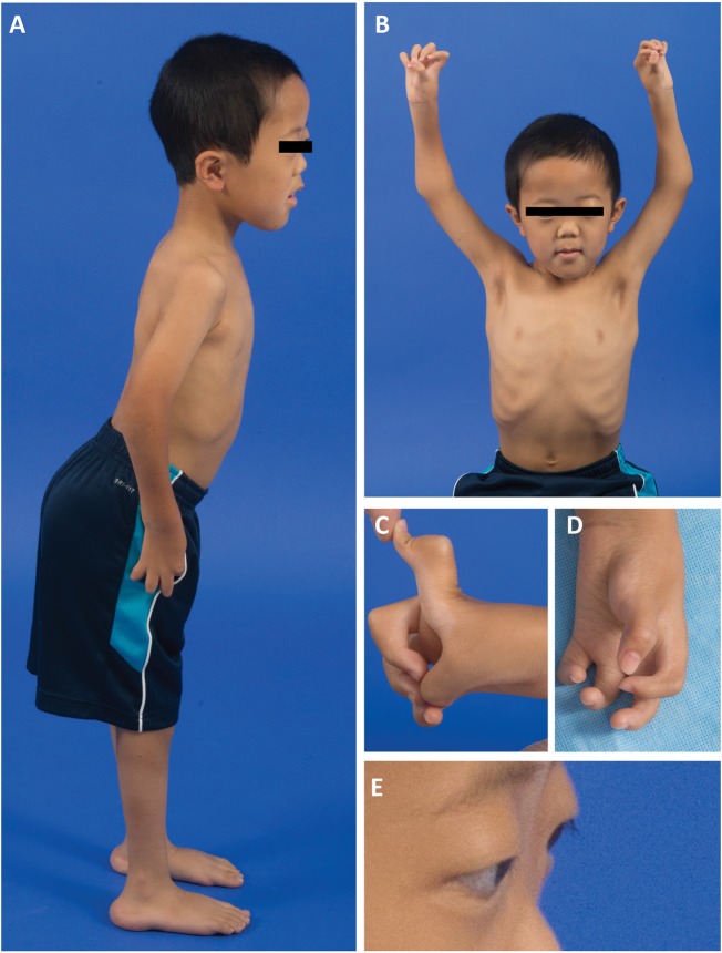
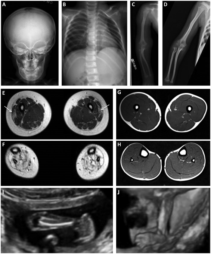
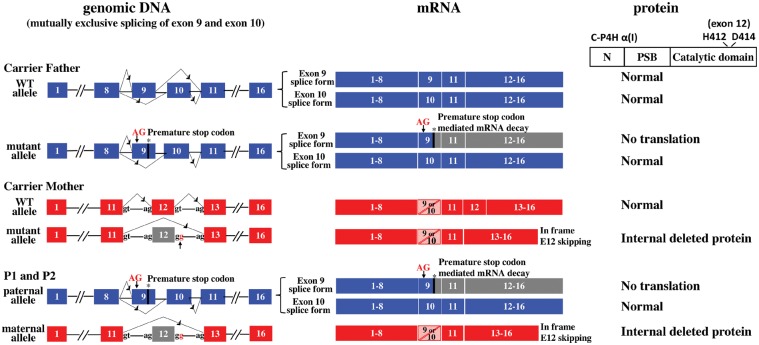
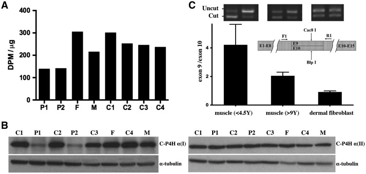
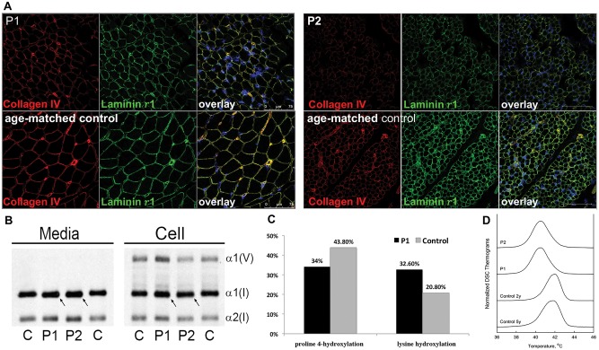
Collagen prolyl 4-hydroxylases (C-P4Hs) play a central role in the formation and stabilization of the triple helical domain of collagens. P4HA1 encodes the catalytic α(I) subunit of the main C-P4H isoenzyme (C-P4H-I). We now report human bi-allelic P4HA1 mutations in a family with a congenital-onset disorder of connective tissue, manifesting as early-onset joint hypermobility, joint contractures, muscle weakness and bone dysplasia as well as high myopia, with evidence of clinical improvement of motor function over time in the surviving patient. Similar to P4ha1 null mice, which die prenatally, the muscle tissue from P1 and P2 was found to have reduced collagen IV immunoreactivity at the muscle basement membrane. Patients were compound heterozygous for frameshift and splice site mutations leading to reduced, but not absent, P4HA1 protein level and C-P4H activity in dermal fibroblasts compared to age-matched control samples. Differential scanning calorimetry revealed reduced thermal stability of collagen in patient-derived dermal fibroblasts versus age-matched control samples. Mutations affecting the family of C-P4Hs, and in particular C-P4H-I, should be considered in patients presenting with congenital connective tissue/myopathy overlap disorders with joint hypermobility, contractures, mild skeletal dysplasia and high myopia.


Figures

Figure 1
Figure 1
Clinical features of the proband (P1) at age 6½ years. (A) P1 with a lordotic stance, flat feet and elbow contractures. (B) Note macrocephaly (> 90th % for age), a broad nasal bridge, a flat midface, frontal bossing, and an asymmetric pectus carinatum (left > right). (C and D) Significant webbing between fingers limiting finger extension, joint hypermobility and absent flexor palmar creases of the hands. (E) Evidence of ocular proptosis and ptosis.
Figure 2
Figure 2
(A–C) Skeletal survey performed at the age 3 years in P1 revealing osteopenia. (A) Anterior-posterior (AP) view of skull with evidence of Wormian bones and a flattened AP diameter of the facial bones. (B) Ribs (AP, oblique) appear thin and slightly angulated. (C) On AP view of the long bones of the forearm, there is evidence of angulation of the right radius and ulna, and the distance between the proximal radius and distal humerus appears possibly increased. (D) Skeletal survey repeated at age 6½ years demonstrating improved osteopenia on AP view of the long bones of the forearm. (E-H) Muscle MRI. Lower extremity muscle MRI performed at age 6½ years in P1 (E and F) revealing abnormal signaling in the vastus lateralis bilaterally (E; arrows) and some atrophy of the quadriceps group but otherwise more normal-appearing muscle above the knee. In contrast, all of the muscles below the knee show severely abnormal signaling, suggestive of replacement of the muscle with connective tissue and/or adipose tissue (F). Muscle MRI in a patient without muscle disease demonstrating normal size and signaling of the muscles above the knee (G) and the muscles below the knee (H). (I and J) Fetal ultrasound images of P2. (I) At 19 weeks gestational age there was evidence of talipes equinovarus. (J) A follow-up fetal ultrasound performed at 21 weeks gestational age revealed continued abnormal posturing of the feet.
Figure 3
Figure 3
Schematic of P4HA1 mutations and consequence at mRNA and protein level. Paternal wild type and mutant allele are shown on top (blue): Each allele has mutually exclusive exon 9 and exon 10 splice forms. The two-base pair insertion in exon 9 [c.1323_1324insAG; p.Arg362Glyfs*9; NM_001017962.2] leads to a premature stop of the exon 9 splice form, as a result this transcript is eliminated by NMD and no protein is translated. Due to the mutually exclusive alternative splicing of exon 9 and 10, the exon 10 containing splice form remains wild type sequence. Maternal wild type and mutant allele are shown in the middle (red): the splice donor site mutation [c.1543 + 2 T > G; p.Ala418_Arg434del; NM_001017962.2)] causes exon 12 skipping and an internally deleted protein. P1 and P2 inherited the paternal and maternal mutant alleles (bottom), as a result, the only normal C-P4H α (I) is translated from the exon 10 splice form of the mutant paternal allele.
Figure 4
Figure 4
(A) Total C-P4H activity was reduced in cultured fibroblasts from P1 and P2 compared to age-matched controls (C1 and C2). Activities in both carrier father (F) and carrier mother (M) were within normal range, although it was at the lower end of the range in the carrier mother. (B) Western blot analysis with α-tubulin loading control. C-P4H α (I) protein is reduced in patients-derived fibroblasts compared to age-matched controls (C1 and C2). There is no up-regulation of C-P4H α (II) protein in cultured fibroblasts from P1, P2 and their parents compared to controls. (C) Ratio of exon 9 versus exon 10 splice form in muscle tissue and cultured dermal fibroblasts from individuals of different ages as determined by RT-PCR followed with Cac8I (specific for exon 9) or BlpI (specific for exon 10) digestion. The exon 9 splice form is the major form expressed in muscle tissue, the average ratio of exon 9/exon 10 is 4.22 (SEM = 0.59) in younger individuals (<4.5 years), the average ratio of exon 9/exon 10 is 2.05 (SEM = 0.09) in older individuals (9 years to adult). The expression levels of exon 9 and exon 10 splice form are relatively equal in dermal fibroblasts at all ages tested (ranges from 8 months to adult, average ratio of exon 9/exon 10 = 0.92, SEM = 0.03).
Figure 5
Figure 5
(A) In muscle biopsies of affected siblings P1 and P2, the basement membrane stains continuously on immunohistochemistry, but both collagen IV and laminin γ1 staining were reduced compared to age-matched controls. (B) Analysis of type I collagen protein shows slightly increased migration of media and cell layer collagens in P1 and P2 compared to control (C). (C) Collagen proline 4-hydroxylation levels were decreased from 43.8% in control (C) to 34% in proband (P1), consistent with the hypomorphic nature of the mutations; while lysine hydroxylation was increased from 20.8% in control to 32.6% in P1. (D) Differential scanning calorimetry (DSC) revealed apparent melting temperature Tm (temperature at the denaturation peak maximum) that was reduced ∼1.3 °C in P1 and P2 (Tm= 40.5 °C in both) compared to 2 y.o, (Tm= 41.9 °C) and 5y.o. normal controls (Tm= 41.7 °C). The broadening and trailing edge of the P1 and P2 denaturation thermograms suggested the presence of a heterogenous population of type I collagen molecules with different thermal stabilities.

References

    1. Mao J.R., Bristow J. (2001) The Ehlers-Danlos syndrome: on beyond collagens. J. Clin. Invest., 107, 1063–1069. - PMC - PubMed
    1. Kivirikko K.I., Pihlajaniemi T. (1998) Collagen hydroxylases and the protein disulfide isomerase subunit of prolyl 4-hydroxylases. Adv. Enzymol Relat. Areas Mol. Biol., 72, 325–398. - PubMed
    1. Myllyharju J. (2003) Prolyl 4-hydroxylases, the key enzymes of collagen biosynthesis. Matrix Biol., 22, 15–24. - PubMed
    1. Myllyharju J., Kivirikko K.I. (2004) Collagens, modifying enzymes and their mutations in humans, flies and worms. Trends Genet., 20, 33–43. - PubMed
    1. Helaakoski T., Vuori K., Myllyla R., Kivirikko K.I., Pihlajaniemi T. (1989) Molecular cloning of the alpha-subunit of human prolyl 4-hydroxylase: the complete cDNA-derived amino acid sequence and evidence for alternative splicing of RNA transcripts. Proc. Natl. Acad. Sci. USA, 86, 4392–4396. - PMC - PubMed

Publication types