

Laboratory x-ray micro-computed tomography: a user guideline for biological samples
- PMID: 28419369
- PMCID: PMC5449646
- DOI: 10.1093/gigascience/gix027
Laboratory x-ray micro-computed tomography: a user guideline for biological samples
Abstract
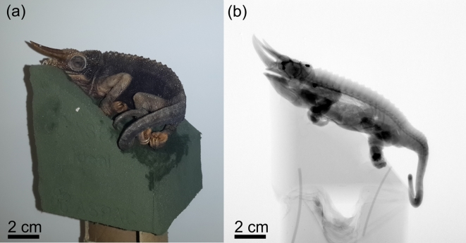
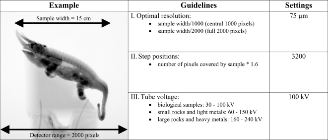
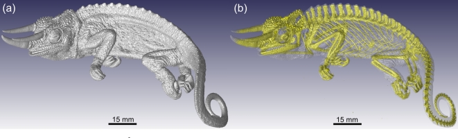
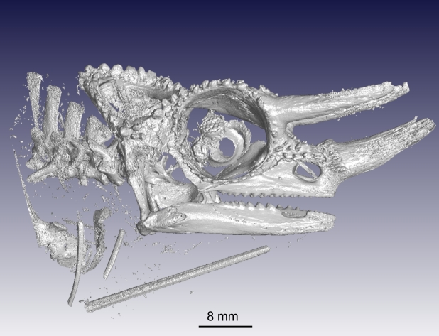
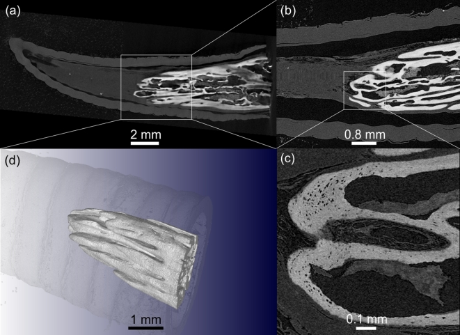
Laboratory x-ray micro-computed tomography (micro-CT) is a fast-growing method in scientific research applications that allows for non-destructive imaging of morphological structures. This paper provides an easily operated "how to" guide for new potential users and describes the various steps required for successful planning of research projects that involve micro-CT. Background information on micro-CT is provided, followed by relevant setup, scanning, reconstructing, and visualization methods and considerations. Throughout the guide, a Jackson's chameleon specimen, which was scanned at different settings, is used as an interactive example. The ultimate aim of this paper is make new users familiar with the concepts and applications of micro-CT in an attempt to promote its use in future scientific studies.
Keywords: 3D imaging; micro-computed tomography; nano-computed tomography; non-destructive analysis; x-ray tomography.
© The Authors 2017. Published by Oxford University Press.
Figures
References
-
- Kalender WA. X-ray computed tomography. Phys Med Biol 2006;51(13):29–43. - PubMed
-
- Brunke O. High-resolution CT-based defect analysis and dimensional measurement. Insight-Non-Destruct Testing Condition Monitor 2010;52(2):91–3.
-
- Singhal A, Grande JC, Zhou Z. Micro/nano CT for visualization of internal structures. Microscopy Today 2013;21(02):16–22.
-
- Schoeman L, Williams P, du Plessis A, et al. . X-ray micro-computed tomography (μCT) for non-destructive characterisation of food microstructure. Trends Food Sci Technol 2016;47:10–24.
-
- Cnudde V, Boone MN. High-resolution x-ray computed tomography in geosciences: a review of the current technology and applications. Earth-Sci Rev 2013;123:1–17.
Publication types
MeSH terms
LinkOut - more resources
Full Text Sources
Other Literature Sources
