

Muscle cell communication in development and repair
- PMID: 28419894
- PMCID: PMC5641474
- DOI: 10.1016/j.coph.2017.03.008
Muscle cell communication in development and repair
Abstract
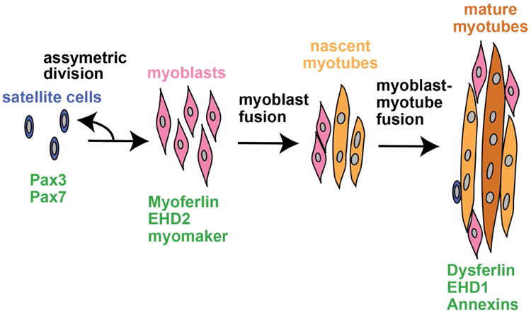
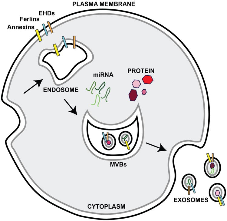
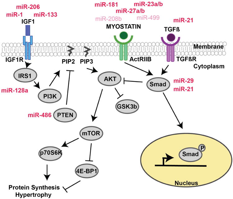
Under basal conditions, postnatal skeletal muscle displays little cell turnover. With injury, muscle initiates a rapid repair response to reseal damaged membrane, reactivating many developmental pathways to facilitate muscle regeneration and prevent tissue loss. Muscle precursor cells become activated accompanied by differentiation and fusion during both muscle growth and regeneration; inter-cellular communication is required for successful completion of these processes. Cellular communication is mediated by lipids, fusogenic membrane proteins, and exosomes. Muscle-derived exosomes carry proteins and micro RNAs as cargo. Secreted factors such as IGF-1, TGFβ, and myostatin are also released by muscle cells providing local signaling cues to modulate muscle fusion and regeneration. Proteins that regulate myoblast fusion also participate in membrane repair and regeneration. Here we will review methods of muscle cell communication focusing on proteins that mediate membrane fusion, exosomes, and autocrine factors.
Copyright © 2017 Elsevier Ltd. All rights reserved.
Conflict of interest statement
Figures
Similar articles
-
Exosomes from differentiating human skeletal muscle cells trigger myogenesis of stem cells and provide biochemical cues for skeletal muscle regeneration.J Control Release. 2016 Jan 28;222:107-15. doi: 10.1016/j.jconrel.2015.12.018. Epub 2015 Dec 14. J Control Release. 2016. PMID: 26699421
-
Mesenchymal-stem-cell-derived exosomes accelerate skeletal muscle regeneration.FEBS Lett. 2015 May 8;589(11):1257-65. doi: 10.1016/j.febslet.2015.03.031. Epub 2015 Apr 8. FEBS Lett. 2015. PMID: 25862500
-
Membrane fusion in muscle development and repair.Semin Cell Dev Biol. 2015 Sep;45:48-56. doi: 10.1016/j.semcdb.2015.10.026. Epub 2015 Oct 30. Semin Cell Dev Biol. 2015. PMID: 26537430 Free PMC article. Review.
-
Exosomes: small vesicles participating in intercellular communication.Int J Biochem Cell Biol. 2012 Jan;44(1):11-5. doi: 10.1016/j.biocel.2011.10.005. Epub 2011 Oct 19. Int J Biochem Cell Biol. 2012. PMID: 22024155 Review.
-
Characteristics and Roles of Exosomes in Cardiovascular Disease.DNA Cell Biol. 2017 Mar;36(3):202-211. doi: 10.1089/dna.2016.3496. Epub 2017 Jan 23. DNA Cell Biol. 2017. PMID: 28112546 Review.
Cited by
-
Reciprocal communication between FAPs and muscle cells via distinct extracellular vesicle miRNAs in muscle regeneration.Proc Natl Acad Sci U S A. 2024 Mar 12;121(11):e2316544121. doi: 10.1073/pnas.2316544121. Epub 2024 Mar 5. Proc Natl Acad Sci U S A. 2024. PMID: 38442155 Free PMC article.
-
The Neuromuscular Disorder Mediated by Extracellular Vesicles in Amyotrophic Lateral Sclerosis.Curr Issues Mol Biol. 2024 Jun 14;46(6):5999-6017. doi: 10.3390/cimb46060358. Curr Issues Mol Biol. 2024. PMID: 38921029 Free PMC article. Review.
-
Enhanced secretion of promyogenic exosomes by quiescent muscle cells.Front Cell Dev Biol. 2024 Jul 23;12:1381357. doi: 10.3389/fcell.2024.1381357. eCollection 2024. Front Cell Dev Biol. 2024. PMID: 39108837 Free PMC article.
-
RNA-Seq identifies genes whose proteins are transformative in the differentiation of cytotrophoblast to syncytiotrophoblast, in human primary villous and BeWo trophoblasts.Sci Rep. 2018 Mar 23;8(1):5142. doi: 10.1038/s41598-018-23379-2. Sci Rep. 2018. PMID: 29572450 Free PMC article.
-
Dynamic Aging: Channeled Through Microenvironment.Front Physiol. 2021 Jul 21;12:702276. doi: 10.3389/fphys.2021.702276. eCollection 2021. Front Physiol. 2021. PMID: 34366891 Free PMC article. Review.
References
-
- Janssen I, Heymsfield SB, Wang ZM, Ross R. Skeletal muscle mass and distribution in 468 men and women aged 18-88 yr. J Appl Physiol (1985) 2000;89:81–88. - PubMed
-
- Grounds MD. The need to more precisely define aspects of skeletal muscle regeneration. Int J Biochem Cell Biol. 2014;56:56–65. - PubMed
-
- Pediconi MF, Donoso P, Hidalgo C, Barrantes FJ. Lipid composition of purified transverse tubule membranes isolated from amphibian skeletal muscle. Biochim Biophys Acta. 1987;921:398–404. - PubMed
-
- Lau YH, Caswell AH, Brunschwig JP, Baerwald R, Garcia M. Lipid analysis and freeze-fracture studies on isolated transverse tubules and sarcoplasmic reticulum subfractions of skeletal muscle. J Biol Chem. 1979;254:540–546. - PubMed
Publication types
MeSH terms
Substances
Grants and funding
LinkOut - more resources
Full Text Sources
Other Literature Sources
Miscellaneous
