Review
. 2017 Apr 16;9(4):81.
doi: 10.3390/v9040081.

Mechanisms of LTR-Retroelement Transposition: Lessons from Drosophila melanogaster

Affiliations
Review

Mechanisms of LTR-Retroelement Transposition: Lessons from Drosophila melanogaster

Lidia Nefedova et al. Viruses. .

Abstract

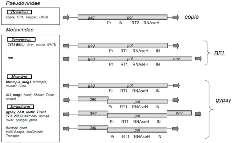
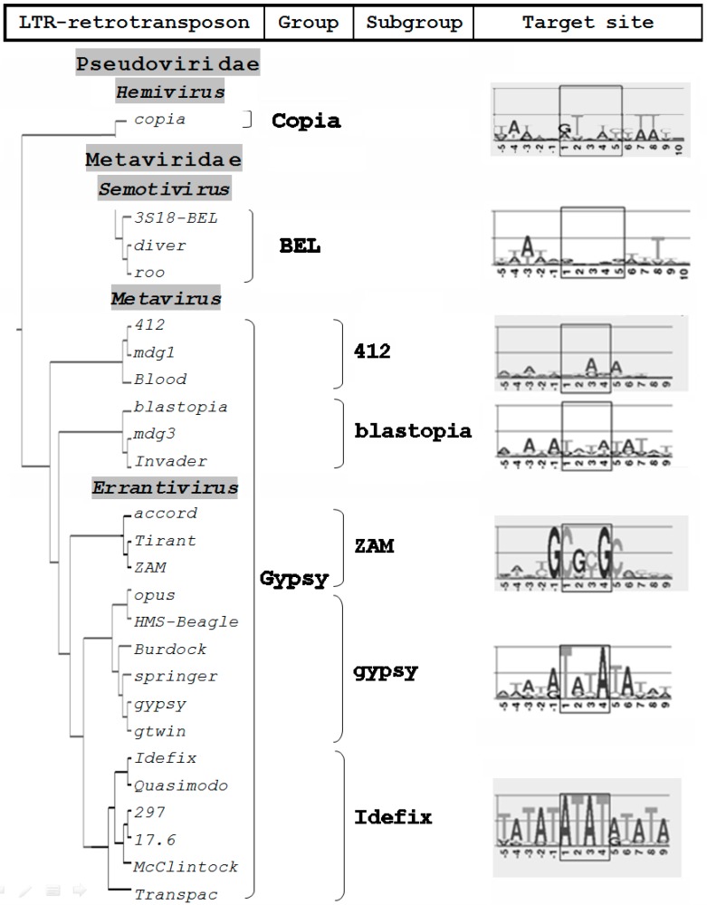
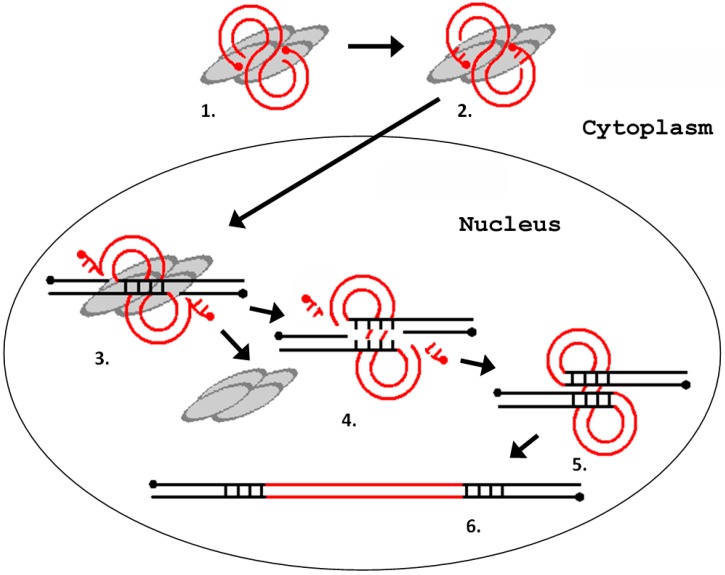
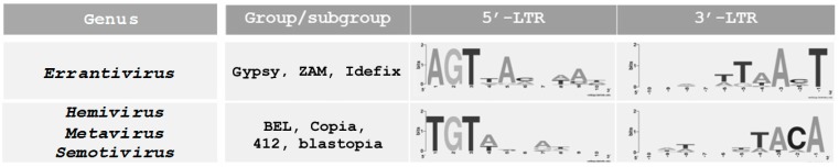
Long terminal repeat (LTR) retrotransposons occupy a special place among all mobile genetic element families. The structure of LTR retrotransposons that have three open reading frames is identical to DNA forms of retroviruses that are integrated into the host genome. Several lines of evidence suggest that LTR retrotransposons share a common ancestry with retroviruses and thus are highly relevant to understanding mechanisms of transposition. Drosophila melanogaster is an exceptionally convenient model for studying the mechanisms of retrotransposon movement because many such elements in its genome are transpositionally active. Moreover, two LTRretrotransposons of D. melanogaster, gypsy and ZAM, have been found to have infectious properties and have been classified as errantiviruses. Despite numerous studies focusing on retroviral integration process, there is still no clear understanding of integration specificity in a target site. Most LTR retrotransposons non-specifically integrate into a target site. Site-specificity of integration at vertebrate retroviruses is rather relative. At the same time, sequence-specific integration is the exclusive property of errantiviruses and their derivatives with two open reading frames. The possible basis for the errantivirus integration specificity is discussed in the present review.

Keywords: Drosophila; LTR‐retrotransposon; errantivirus; retrovirus; transposition.


Conflict of interest statement

The authors declare no conflict of interest. The founding sponsors had no role in the design of the study; in the collection, analyses, or interpretation of data; in the writing of the manuscript, and in the decision to publish the results.

Figures

Figure 1
Figure 1
Structural organization and classification of Drosophila melanogaster long terminal repeat (LTR) retrotransposons. As shown: open reading frames (gag, pol, env) and the Pol domains (Pr, protease; RT1 and RT2, reverse transcriptase types 1 and 2; IN, integrase). The arrows indicate LTRs. LTR retrotransposons are distributed in groups and genera according to the phylogenetic analysis conducted in [9]. The LTR retrotransposon families introduced by the International Committee on Taxonomy of Viruses (ICTV) in Metaviridae and Pseudoviridae are highlighted in bold.
Figure 2
Figure 2
Integration sites of D. melanogaster LTR retrotransposons. A phylogenetic tree construction is based on a comparison of the amino acid sequences of the integrases of D. melanogaster LTR retrotransposons [25]. Visualization of the target site duplication was made using WEBLOGO (version 3) [29].
Figure 3
Figure 3
Phylogenetic tree of the 5’-untranlsated region (5′-UTR) repeat module (102 base pair; bp) in Tirant of D. simulans (subfamilies S and C) and D. melanogaster (subfamilies Tirant and Tirant_het) and sequence identity matrix (%).
Figure 4
Figure 4
Schematic representation of the process of integration of a retrovirus (LTR retrotransposon) into the host genome. (1) interaction of integrase with a blunt-ended DNA substrate (other proteins are not shown); (2) removal of two terminal nucleotides from the 3′-ends of the DNA substrate (3′-end processing); (3) cleavage of the integration site; (4) removal of unpaired nucleotides at the 5′ ends of the DNA substrate; (5) filling-in of the gaps of the target DNA and ligation of discontinuities; and (6) repeating of the provirus integration site. The gray ovals represent integrase monomers. The red lines represent viral DNA, and the black lines represent chromosomal DNA. The dots indicate 5′-ends of the DNA.
Figure 5
Figure 5
Multiple alignment of the 5′- and 3′-terminal sequences of the D. melanogaster LTR retrotransposons. The LTR retrotransposons of D. melanogaster can be divided into two groups depending on the composition of the end sequences [25]. Visualization performed using WEBLOGO [29].

Similar articles

Cited by

References

    1. De Koning A.P., Gu W., Castoe T.A., Batzer M.A., Pollock D.D. Repetitive elements may comprise over two-thirds of the human genome. PLoS Genet. 2011;7:e1002384. doi: 10.1371/journal.pgen.1002384. - DOI - PMC - PubMed
    1. Bergman C.M., Quesneville H. Discovering and detecting transposable elements in genome sequences. Brief. Bioinform. 2007;8:382–392. doi: 10.1093/bib/bbm048. - DOI - PubMed
    1. Petrov D.A., Fiston-Lavier A.S., Lipatov M., Lenkov K., González J. Population genomics of transposable elements in Drosophila melanogaster. Mol. Biol. Evol. 2011;28:633–644. doi: 10.1093/molbev/msq337. - DOI - PMC - PubMed
    1. Kim A., Terzian С., Santamaria P., Pélisson A., Prud’homme N., Bucheton A. Retroviruses in vertebrates: The gypsy retrotransposon is apparently an infectious retrovirus of Drosophila melanogaster. Proc. Natl. Acad. Sci. USA. 1994;91:1285–1289. doi: 10.1073/pnas.91.4.1285. - DOI - PMC - PubMed
    1. Leblanc P., Desset S., Giorgi F., Taddei A.R., Fausto A.M., Mazzini M., Dastugue B., Vaury C. Life cycle of an endogenous retrovirus, ZAM, in Drosophila melanogaster. J. Virol. 2000;74:10658–10669. doi: 10.1128/JVI.74.22.10658-10669.2000. - DOI - PMC - PubMed

Publication types

LinkOut - more resources