. 2017 Apr 17;18(4):834.
doi: 10.3390/ijms18040834.

Beneficial Effects of Melatonin on the In Vitro Maturation of Sheep Oocytes and Its Relation to Melatonin Receptors

Affiliations

Beneficial Effects of Melatonin on the In Vitro Maturation of Sheep Oocytes and Its Relation to Melatonin Receptors

Xiuzhi Tian et al. Int J Mol Sci. .

Abstract

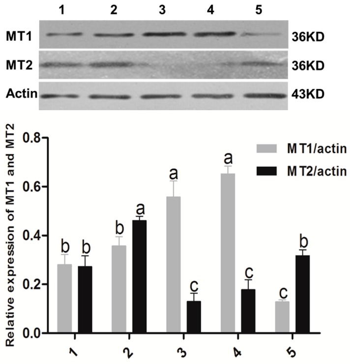
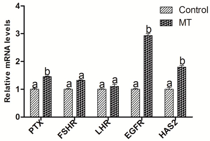
(1) Background: The binding sites of melatonin, as a multifunctional molecule, have been identified in human, porcine, and bovine samples. However, the binding sites and mechanisms of melatonin have not been reported in sheep; (2) Methods: Cumulus-oocyte complexes (COCs) were cultured in TCM-199 supplemented with melatonin at concentrations of 0, 10-3, 10-5, 10-7, 10-9, and 10-11 M. Melatonin receptors (MT1 and MT2) were evaluated via immunofluorescence and Western blot. The effects of melatonin on cumulus cell expansion, nuclear maturation, embryo development, and related gene (GDF9, DNMT1, PTX3, HAS2, and EGFR) expression were investigated. The level of cyclic adenosine monophosphate (cAMP) and cyclic guanosine monophosphate (cGMP) were evaluated in oocytes and cumulus, respectively; (3) Results: Both MT1 and MT2 were expressed in oocytes, cumulus cells, and granulosa cells. Melatonin with a concentration of 10-7 M significantly enhanced the rates of nuclear maturation, cumulus cells expansion, cleavage, and blastocyst. Melatonin enhanced the expression of BMP15 in oocytes and of PTX3, HAS2, and EGFR in cumulus cells. Melatonin decreased the cAMP level of oocytes but enhanced the cGMP level in oocytes and cumulus cells; (4) Conclusion: The higher presence of MT1 in GV cumulus cells and the beneficial effects of melatonin indicated that its roles in regulating sheep oocyte maturation may be mediated mainly by the MT1 receptor.

Keywords: MT1/MT2; meioctic maturation; melatonin; oocyte; sheep.


Conflict of interest statement

The authors declare no conflict of interest.

Figures

Figure 1
Figure 1
Assayed the melatonin receptor with immunofluorescence. The cellular localizations of MT1/MT2 were identified by confocal analysis. Sheep COCs were incubated with the MT1/MT2 antibody (red) followed by Alexa-488-conjugated donkey anti-goat IgG (green). (A) images of MT1 receptor; (B) images of MT2 receptor. GV: germinal vesicle stage, MII: metaphase II stage. Scale bar = 50 µm.
Figure 1
Figure 1
Assayed the melatonin receptor with immunofluorescence. The cellular localizations of MT1/MT2 were identified by confocal analysis. Sheep COCs were incubated with the MT1/MT2 antibody (red) followed by Alexa-488-conjugated donkey anti-goat IgG (green). (A) images of MT1 receptor; (B) images of MT2 receptor. GV: germinal vesicle stage, MII: metaphase II stage. Scale bar = 50 µm.
Figure 2
Figure 2
The expression of MT1 and MT2 identified with Western blot method. 1. Oocyte in GV stage; 2. Oocyte in MII stage; 3. Granulosa cells; 4. Cumulus cells in GV stage; 5. Cumulus cells in MII stage. The superscript different letters (a–c) represent a significant difference in the same column (p < 0.05).
Figure 3
Figure 3
Effects of melatonin on cumulus cell expansion and oocyte nuclear maturation. Maturation rate of oocytes treated with melatonin (0, 10−3, 10−5, 10−7, 10−9, and 10−11 M) for 24 h, cumulus cell expansion, and polar body extrusion were analyzed, respectively. (A) No expansion percentage; (B) Partial expansion percentage; (C) Complete expansion percentage; (D) Polar extrusion percentage. Data are expressed as percentage ± SEM from six independent experiments (>30 oocytes per treatment per experiment). Bars with different letters (a–c) represent significantly different (p < 0.05).
Figure 4
Figure 4
Effect of melatonin on the development of parthenogenetic activation sheep oocytes. Embryos were acquired by parthenogenetic activation oocytes that were cultured in the maturation medium contained different concentrations of melatonin. (A) cleavage rate and blastocyst rate; (B) cell number/blastocyst. The superscript different letters (a–d) represent a significant difference in the same column (p < 0.05).
Figure 5
Figure 5
Effects of 10−7 M melatonin on the gene expression in sheep oocytes. The genes include the oocyte-secreted factors (GDF9 and BMP15) and DNA methyltransferase 1 (DNMT1). The letters a and b identify statistically significant differences (p < 0.05).
Figure 6
Figure 6
Effects of 10−7 M melatonin on the gene expression of cumulus cells expansion. The genes include PTX3, HAS2, LHR, FSHR, and EGFR. The letters (a,b) identify statistically significant differences (p < 0.05).
Figure 7
Figure 7
Effects of melatonin receptor antagonist luzindole (106 M) on the in vitro maturation of sheep oocytes. (A) PB1 extrusion rate and cleavage rate; (B) Blastocyst rate; (C) Hatched blastocyst rate; (D) Cell number/blastocyst. The numbers of cultured oocytes for each group varied from 200 to 250, respectively. Different superscript letters (a,b) in each column identify statistical significant differences (p < 0.05).
Figure 8
Figure 8
Intracellular cAMP concentration of oocytes and cumulus cells in different treatments. (A) The concentration of cAMP per oocyte or cumulus cell; (B) The concentration of cGMP per oocyte or cumulus cell. Data are from five independent repeats and are shown as mean ± SEM. The superscript different letters (a,b) represent a significant difference in the same column compared to the control (p < 0.05).
Figure 9
Figure 9
Schematic representation of melatonin roles on promoting oocytes maturation. PDE: phosphodiesterase; cGMP: cyclic guanosine monophosphate; cAMP: cyclic adenosine monophosphate. Red arrow: promoting action; blue arrow: gene expression improved; green arrow: improved gene expression resulted in cumulus cells expansion; red T bar: inhibiting effect.

Similar articles

Cited by

References

    1. Dobrinsky J.R., Johnson L.A., Rath D. Development of a culture medium (BECM-3) for porcine embryos: Effects of bovine serum albumin and fetal bovine serum on embryo development. Biol. Reprod. 1996;55:1069–1074. doi: 10.1095/biolreprod55.5.1069. - DOI - PubMed
    1. Adriaens I., Jacquet P., Cortvrindt R., Janssen K., Smitz J. Melatonin has dose-dependent effects on folliculogenesis, oocyte maturation capacity and steroidogenesis. Toxicology. 2006;228:333–343. doi: 10.1016/j.tox.2006.09.018. - DOI - PubMed
    1. Kang J.T., Koo O.J., Kwon D.K., Park H.J., Jang G., Kang S.K., Lee B.C. Effects of melatonin on in vitro maturation of porcine oocyte and expression of melatonin receptor RNA in cumulus and granulosa cells. J. Pineal Res. 2009;46:22–28. doi: 10.1111/j.1600-079X.2008.00602.x. - DOI - PubMed
    1. Shi J., Tian X., Zhou G., Wang L., Gao C., Zhu S., Zeng S., Tian J., Liu G. Melatonin exists in porcine follicular fluid and improves in vitro maturation and parthenogenetic development of porcine oocytes. J. Pineal Res. 2009;47:318–323. doi: 10.1111/j.1600-079X.2009.00717.x. - DOI - PubMed
    1. Papis K., Poleszczuk O., Wenta-Muchalska E., Modlinski J.A. Melatonin effect on bovine embryo development in vitro in relation to oxygen concentration. J. Pineal Res. 2007;43:321–326. doi: 10.1111/j.1600-079X.2007.00479.x. - DOI - PubMed