. 2017 Apr 17;18(4):846.
doi: 10.3390/ijms18040846.

The Angiogenesis Inhibitor ALS-L1023 from Lemon-Balm Leaves Attenuates High-Fat Diet-Induced Nonalcoholic Fatty Liver Disease through Regulating the Visceral Adipose-Tissue Function

Affiliations

The Angiogenesis Inhibitor ALS-L1023 from Lemon-Balm Leaves Attenuates High-Fat Diet-Induced Nonalcoholic Fatty Liver Disease through Regulating the Visceral Adipose-Tissue Function

Jeongjun Kim et al. Int J Mol Sci. .

Abstract

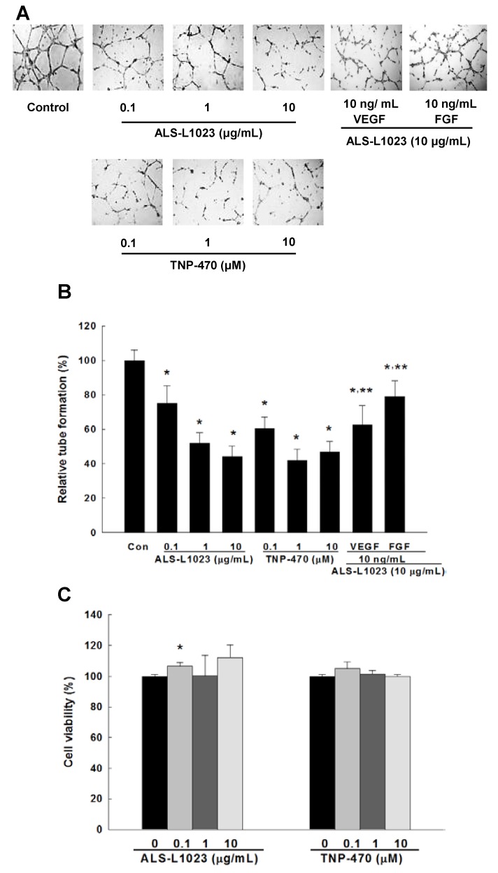
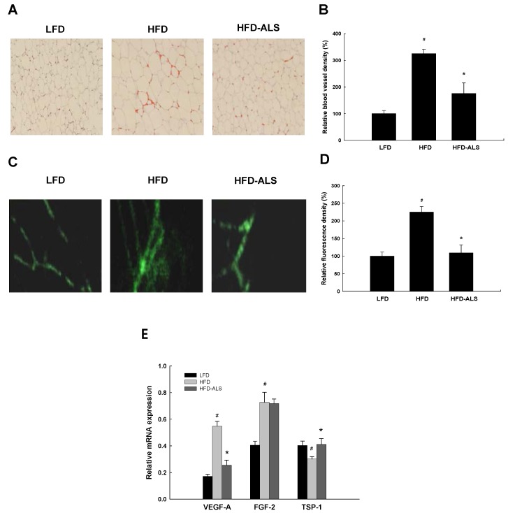
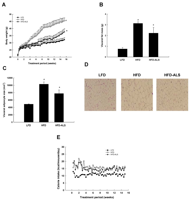
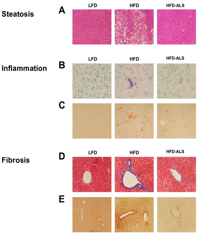
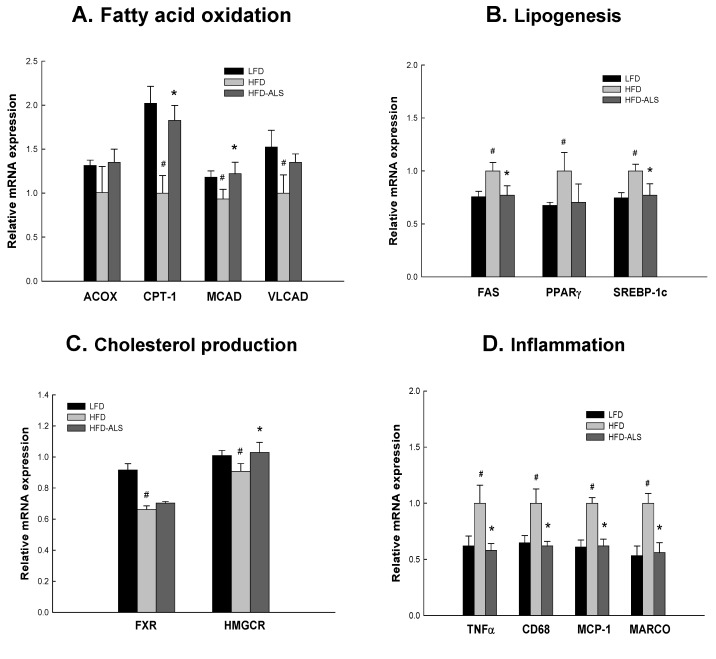
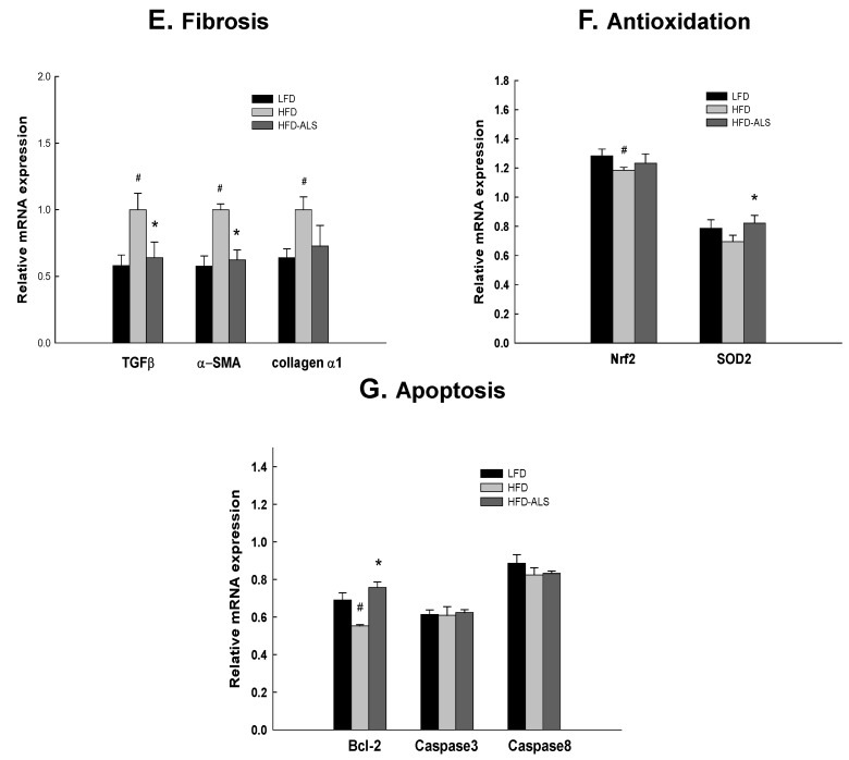
Similar to neoplastic tissues, growth and development of adipose tissue are thought to be angiogenesis-dependent. Since visceral adipose tissue (VAT) is associated with development and progression of nonalcoholic fatty liver disease (NAFLD), we hypothesized that angiogenesis inhibition would attenuate obesity-induced NAFLD. We fed C57BL/6J mice a low-fat diet (LFD, chow 10% kcal fat), a high-fat diet (HFD, 45% kcal fat) or HFD supplemented with the lemon-balm extract ALS-L1023 (HFD-ALS) for 15 weeks. ALS-L1023 reduced endothelial cell-tube formation in vitro. HFD increased VAT angiogenesis and induced weight gains including body weight, VAT mass and visceral adipocyte size compared with LFD. However, HFD-ALS led to weight reductions without affecting calorie intake compared with HFD. HFD-ALS also reduced serum ALT and AST levels and improved lipid metabolism. HFD-ALS suppressed steatosis, infiltration of inflammatory cells, and accumulation of collagen in livers. HFD-ALS modulated hepatic expression of genes involved in lipid metabolism, inflammation, fibrosis, antioxidation, and apoptosis. Concomitantly, analysis of VAT function revealed that HFD-ALS led to fewer CD68-positive macrophage numbers and lower expression of inflammatory cytokines compared with HFD. Our findings show that the anti-angiogenic herbal extract ALS-L1023 attenuates NAFLD by targeting VAT during obesity, suggesting that angiogenesis inhibitors could aid in the treatment and prevention of obesity-induced human NAFLD.

Keywords: Melissa officinalis; herbal medicine; visceral adipose inflammation; visceral obesity.


Conflict of interest statement

The authors declare no conflict of interest.

Figures

Figure 1
Figure 1
Inhibitory effects of ALS-L1023 on endothelial cell-tube formation in vitro. (A) tube formation of human endothelial cells. Cells were plated in Matrigel-coated wells and incubated with medium containing ALS-L1023, TNP-470, ALS-L1023 plus vascular endothelial growth factor (VEGF) or ALS-L1023 plus fibroblast growth factor (FGF). After incubation, capillary-like tube formation was photographed (original magnification ×100); (B) inhibition of tube formation; (C) human endothelial cell viability by 2,3-bis[2-methoxy-4-nitro-5-sulfophenyl]-2H-tetrazolium-5-carboxanilide disodium salt (XTT) assay. All values are expressed as the mean ± SD (n = 9/group). * p < 0.05 compared with control. ** p < 0.05 compared with ALS-L1023.
Figure 2
Figure 2
Inhibitory effects of ALS-L1023 on VAT vasculature in vivo. Adult male C57BL/6J mice were fed a low-fat diet (LFD), a high-fat diet (HFD) or HFD supplemented with ALS-L1023 (HFD-ALS) for 15 weeks. (A) immunohistochemical detection of vWF-positive cells in the visceral adipose tissue (VAT) (original magnification ×100); (B) inhibition of blood vessel density by ALS-L1023; (C) real-time fibered confocal fluorescence microscopic images of VAT angiogenesis; (D) analysis of fluorescence density in the VAT; (E) the mRNA expression of vascular endothelial growth factor A (VEGF-A), fibroblast growth factor 2 (FGF-2), and thrombospondin-1 (TSP-1) in the VAT. All values are expressed as the mean ± SD (n = 5/group). # p < 0.05 compared with LFD. * p < 0.05 compared with HFD.
Figure 3
Figure 3
Effects of ALS-L1023 on body weight, visceral fat mass, visceral adipocyte size, and food consumption in HFD mice. Adult male C57BL/6J mice were fed a low-fat diet (LFD), a high-fat diet (HFD) or HFD supplemented with ALS-L1023 (HFD-ALS) for 15 weeks. (A) body weights at the end of the treatment period are significantly different between the LFD group and the HFD group (# p < 0.05) and between the HFD group and the group fed a HFD supplemented with ALS-L1023 (* p < 0.05); (B) visceral adipose tissue mass and (C) epididymal adipocyte size; (D) representative hematoxylin-eosin-stained sections (5 µm thick) of epididymal adipose tissues are shown (original magnification ×100); (E) effects of ALS-L1023 on food intake. All values are expressed as the mean ± SD (n = 8/group). # p < 0.05 compared with LFD. * p < 0.05 compared with HFD.
Figure 4
Figure 4
Changes in circulating ALT, AST and lipid levels in HFD mice. Adult male C57BL/6J mice were fed a low-fat diet (LFD), a high-fat diet (HFD) or HFD supplemented with ALS-L1023 (HFD-ALS) for 15 weeks. Serum levels of (A) alanine aminotransferase (ALT) and (B) aspartate transaminase (AST); serum levels of (C) triglycerides; (D) free fatty acids and (E) total cholesterol. All values are expressed as the mean ± SD (n = 8/group). # p < 0.05 compared with LFD. * p < 0.05 compared with HFD.
Figure 5
Figure 5
Effects of ALS-L1023 on liver histology in HFD mice. Adult male C57BL/6J mice were fed a low-fat diet (LFD), a high-fat diet (HFD) or HFD supplemented with ALS-L1023 (HFD-ALS) for 15 weeks. (A) hematoxylin and eosin-stained sections of liver tissues (original magnification ×100); (B) toluidine blue-stained sections of liver tissues (original magnification ×400); (C) immunohistochemical detection of CD68-positive macrophages in the liver (original magnification ×200); (D) Masson’s trichrome-stained sections of liver tissues (original magnification ×100); (E) liver sections stained with an antibody against α-smooth muscle actin (α-SMA) (original magnification ×200). All values are expressed as the mean ± SD (n = 10/group).
Figure 6
Figure 6
Effects of ALS-L1023 on hepatic gene expression in HFD mice. Adult male C57BL/6J mice were fed a low-fat diet (LFD), a high-fat diet (HFD) or HFD supplemented with ALS-L1023 (HFD-ALS) for 15 weeks. The mRNA expression of (A) fatty acid oxidation, (B) lipogenic, and (C) cholesterol synthesis genes in the liver. The mRNA expression of (D) inflammatory and (E) fibrogenic genes in the liver. The mRNA expression of (F) antioxidation and (G) apoptosis genes in the liver. All values are expressed as the mean ± SD (n = 6/group). # p < 0.05 compared with LFD. * p < 0.05 compared with HFD. ACOX, acyl-CoA oxidase; α-SMA, α-smooth muscle actin; Bcl-2, B cell lymphoma 2; CPT-1, carnitine palmitoyltransferase I; FAS, fatty acid synthase; FXR, farnesoid X receptor; HMGCR, HMG-CoA reductase; ICAM-1, intercellular adhesion molecule-1; MARCO, macrophage receptor with collagenous structure; MCAD, medium-chain acyl-coenzyme A dehydrogenase; MCP-1, monocyte chemoattractant protein 1; Nrf2, nuclear factor E2-related factor 2; PPARγ peroxisome proliferator-activated receptor γ; SOD2, superoxide dismutase 2; SREBP-1c, sterol regulatory element-binding protein 1c; TGFβ, transforming growth factor β; TNFα, tumor necrosis factor α; VCAM-1, vascular cell adhesion molecule; VLCAD, very long-chain acyl-coenzyme A dehydrogenase.
Figure 6
Figure 6
Effects of ALS-L1023 on hepatic gene expression in HFD mice. Adult male C57BL/6J mice were fed a low-fat diet (LFD), a high-fat diet (HFD) or HFD supplemented with ALS-L1023 (HFD-ALS) for 15 weeks. The mRNA expression of (A) fatty acid oxidation, (B) lipogenic, and (C) cholesterol synthesis genes in the liver. The mRNA expression of (D) inflammatory and (E) fibrogenic genes in the liver. The mRNA expression of (F) antioxidation and (G) apoptosis genes in the liver. All values are expressed as the mean ± SD (n = 6/group). # p < 0.05 compared with LFD. * p < 0.05 compared with HFD. ACOX, acyl-CoA oxidase; α-SMA, α-smooth muscle actin; Bcl-2, B cell lymphoma 2; CPT-1, carnitine palmitoyltransferase I; FAS, fatty acid synthase; FXR, farnesoid X receptor; HMGCR, HMG-CoA reductase; ICAM-1, intercellular adhesion molecule-1; MARCO, macrophage receptor with collagenous structure; MCAD, medium-chain acyl-coenzyme A dehydrogenase; MCP-1, monocyte chemoattractant protein 1; Nrf2, nuclear factor E2-related factor 2; PPARγ peroxisome proliferator-activated receptor γ; SOD2, superoxide dismutase 2; SREBP-1c, sterol regulatory element-binding protein 1c; TGFβ, transforming growth factor β; TNFα, tumor necrosis factor α; VCAM-1, vascular cell adhesion molecule; VLCAD, very long-chain acyl-coenzyme A dehydrogenase.
Figure 7
Figure 7
Epididymal adipose tissue inflammation of HFD-ALS mice. Adult male C57BL/6J mice were fed a low-fat diet (LFD), a high-fat diet (HFD) or HFD supplemented with ALS-L1023 (HFD-ALS) for 15 weeks. (A) hematoxylin and eosin-stained sections of epididymal adipose tissues (original magnification ×100); (B) immunohistochemical detection of CD68+ macrophages in epididymal adipose tissues (original magnification ×200); (C) the mRNA expression of inflammatory factors in epididymal adipose tissues. All values are expressed as mean ± SD (n = 6/group)). # p < 0.05 compared with LFD. * p < 0.05 compared with HFD. MCP-1, monocyte chemoattractant protein 1; TNFα, tumor necrosis factor α.

Similar articles

Cited by

References

    1. Folkman J. Tumor angiogenesis. Adv. Cancer Res. 1985;43:175–203. - PubMed
    1. Celletti FL., Waugh J.M., Amabile P.G., Brendolan A., Hilfiker P.R., Dake M.D. Vascular endothelial growth factor enhances atherosclerotic plaque progression. Nat. Med. 2001;7:425–429. doi: 10.1038/86490. - DOI - PubMed
    1. Ferrara N., Davis-Smyth T. The biology of vascular endothelial growth factor. Endocr. Rev. 1997;18:4–25. doi: 10.1210/edrv.18.1.0287. - DOI - PubMed
    1. Crandall D.L., Hausman G.J., Kral J.G. A review of the microcirculation of adipose tissue: Anatomic, metabolic, and angiogenic perspectives. Microcirculation. 1997;4:211–232. doi: 10.3109/10739689709146786. - DOI - PubMed
    1. Hobson B., Denekamp J. Endothelial proliferation Endothelial proliferation in tumours and normal tissues: Continuous labelling studies. Br. J. Cancer. 1984;49:405–413. doi: 10.1038/bjc.1984.66. - DOI - PMC - PubMed

MeSH terms

LinkOut - more resources