Review
. 2017 Apr 17;18(4):843.
doi: 10.3390/ijms18040843.

Melatonin, a Full Service Anti-Cancer Agent: Inhibition of Initiation, Progression and Metastasis

Affiliations
Review

Melatonin, a Full Service Anti-Cancer Agent: Inhibition of Initiation, Progression and Metastasis

Russel J Reiter et al. Int J Mol Sci. .

Abstract

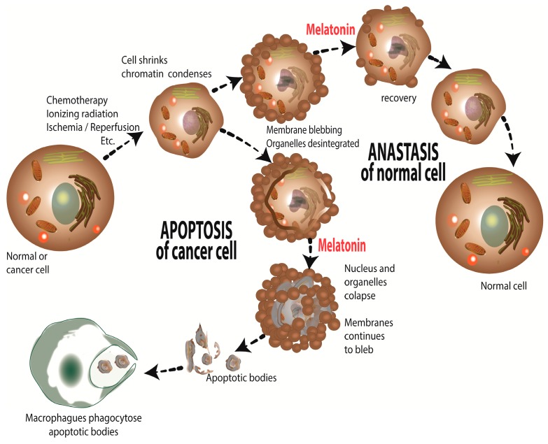
There is highly credible evidence that melatonin mitigates cancer at the initiation, progression and metastasis phases. In many cases, the molecular mechanisms underpinning these inhibitory actions have been proposed. What is rather perplexing, however, is the large number of processes by which melatonin reportedly restrains cancer development and growth. These diverse actions suggest that what is being observed are merely epiphenomena of an underlying more fundamental action of melatonin that remains to be disclosed. Some of the arresting actions of melatonin on cancer are clearly membrane receptor-mediated while others are membrane receptor-independent and involve direct intracellular actions of this ubiquitously-distributed molecule. While the emphasis of melatonin/cancer research has been on the role of the indoleamine in restraining breast cancer, this is changing quickly with many cancer types having been shown to be susceptible to inhibition by melatonin. There are several facets of this research which could have immediate applications at the clinical level. Many studies have shown that melatonin's co-administration improves the sensitivity of cancers to inhibition by conventional drugs. Even more important are the findings that melatonin renders cancers previously totally resistant to treatment sensitive to these same therapies. Melatonin also inhibits molecular processes associated with metastasis by limiting the entrance of cancer cells into the vascular system and preventing them from establishing secondary growths at distant sites. This is of particular importance since cancer metastasis often significantly contributes to death of the patient. Another area that deserves additional consideration is related to the capacity of melatonin in reducing the toxic consequences of anti-cancer drugs while increasing their efficacy. Although this information has been available for more than a decade, it has not been adequately exploited at the clinical level. Even if the only beneficial actions of melatonin in cancer patients are its ability to attenuate acute and long-term drug toxicity, melatonin should be used to improve the physical wellbeing of the patients. The experimental findings, however, suggest that the advantages of using melatonin as a co-treatment with conventional cancer therapies would far exceed improvements in the wellbeing of the patients.

Keywords: angiogenesis; antioxidant; apoptosis; breast; chemotherapy; free radicals; invasion; ionizing radiation; melatonin receptors; molecular mechanisms; prostate.


Conflict of interest statement

The authors declare no conflict of interest.

Figures

Figure 1
Figure 1
Chemical processes involved in ionizing radiation-mediated cellular injury as they relate to an elevated cancer risk. Clustered DNA damage and hydroxyl radical (•OH) generation are direct consequences of ionizing radiation exposure. The resulting damage to DNA in particular, but not exclusively, enhances the possibility of genetic mutations thereby boosting cancer initiation. Mitigating the damage to DNA and other molecules during Earth- and space-related high energy radiation exposure has important implications for health. As summarized in the text, melatonin given supplementally has significant ability to reduce molecular damage due to radiation exposure.
Figure 2
Figure 2
As illustrated here, there are many exogenous and endogenous factors/processes that lead to the intracellular formation of reduced oxygen derivatives. A major contributor to endogenous free radical generation is the escape of electrons (e) from the respiratory complexes of the electron transport chain. This generates the O2 which is quickly dismutated to H2O2 and, via the Haber-Weiss/Fenton reaction, this oxygen derivative is converted to the •OH. Additionally, O2 produced in mitochondria can also diffuse into the cytosol through voltage-dependent anion channels (VDAC, also known as porin). The ROS marked with an X are directly scavenged by melatonin and its metabolites. Also, melatonin stimulates (↑) the activities of the enzymes which metabolize H2O2 to harmless molecules, H2O and O2. The enzymes shown to be either stimulated or protected from damage by melatonin include glutathione peroxidase (GPx), glutathione reductase (GRd) and catalase (CAT). e, electron; H+, hydrogen atom; GSH, reduced glutathione; GSSG, oxidized glutathione; NADP+, nicotinamide adenine dinucleotide phosphate (reduced); NADPH, nicotinamide adenine nucleotide phosphate (oxidized); PRx, thioredoxin peroxidase; SOD2, mitochondrial superoxide dismutase; TM, transition metal; TRx, thioredoxin (oxidized/reduced); TRxP, thioredoxin reductase.
Figure 3
Figure 3
This figure illustrates the likely role of melatonin in influencing L1 expression and DNA damage. During the day (left) which circulating melatonin levels are at their nadir, L1 mRNA causes L1-mediated DNA damage. At night in darkness (center), the elevation of melatonin in the blood acts on the MT1 melatonin receptor to suppress L1 mRNA and ORF1 (open reading frame) protein. By this means, melatonin reduces L1-associated genomic stability thereby reducing the risk of cancer. Contaminating the night with light reduces melatonin levels leading to high L1 mRNA and protein expression causing L1-mediated DNA damage (right).
Figure 4
Figure 4
The figure illustrates some of the processes whereby melatonin may mediate apoptosis of cancer cells. In cancer cells, in contrast to normal cells, melatonin enhances reactive oxygen species (ROS) generation, which lead to cellular death via apoptosis. The text should be consulted for details.
Figure 5
Figure 5
A summary of the theoretical actions of melatonin on constitutive photomorphogenic protein 1 (COP1) as it may relate to cancer. COP1, which we hypothesize is influenced by melatonin, controls the expression of multiple targets in the blue box (check color) thereby impacting tumorigenesis as well as carbohydrate and lipid metabolism. The inclusion of COP9 may potentially relate to melatonin’s antioxidant actions. The presumed direct effect of melatonin on the proteasome is also indicated.
Figure 6
Figure 6
Mechanisms of melatonin’s oncostatic action during prostate cancer progression; these are either independent of the functions of melatonin receptors (left) or mediated via specific interaction of the indoleamine with its receptors (right). Multiple signaling cascades are induced or inactivated in prostate cancer cells upon melatonin treatment, which results in a variety of biological effects of the pineal secretory product. See text for additional details. T, testosterone; P, phosphorylation.
Figure 7
Figure 7
Differential responses of subcutaneously growing human breast tumors to daily intraperitoneally-injected tamoxifen (TAM) alone or concurrently with melatonin (MLT, given in the drinking water) in rats exposed to dim light at night (dLAN). The light:dark cycle was 12:12. dLAN suppressed the nocturnal rise (A) in endogenous melatonin which was restored by the addition of melatonin to the drinking water. dLAN (red dots and line) advanced the latency to recognizable tumor growth (B) compared to that in rats exposed to 12:12 where the night was dark (black dots and line). Also, TAM administered to rats exposed to dLAN failed to inhibit tumor growth (blue dots and lines), i.e., they were resistant to TAM. By comparison, when TAM treatment was instituted in rats that were given melatonin, tumor enlargement was inhibited (green dots and lines), i.e., they became sensitive to TAM. The speculated mechanisms to explain the elevated sensitivity of the tumors to TAM are multiple and are summarized in the text. Figure drawn from the data published by Dauchy et al. [308].
Figure 8
Figure 8
Schematic representation of the multiple mechanisms involved in melatonin-mediated inhibition of cancer metastasis. EMT: Epithelial-mesenchymal transition; ET1: Endothelin-1; GSK3β: Glycogen synthase kinase 3β; HER-2: Human epidermal growth factor receptor-2; MAPK: Mitogen-activated protein kinase; MLCK: Myosin light-chain kinase; MMP-9: Matrix metalloproteinase-9; ROCK1: Rho-associated protein kinase 1; VEGF: Vascular endothelial growth factor.
Figure 9
Figure 9
The multiple means that have been proposed by which melatonin may interfere with the growth of experimental tumors are listed. The blue boxes identify the process that are impacted by melatonin while the green boxes list the potential mechanisms involved. The information in red mentions the synergistic actions of melatonin with radio- or chemotherapies (left) and on the right it is noted that some cancers resistant to therapy can be made sensitive by treatment with melatonin.
Figure 10
Figure 10
Anastasis, a recently-discovered cellular function, describes the recovery a cell undergoes after apoptosis has been initiated but then the stimulus that launched the apoptosis process is withdrawn. As illustrated here, based on what is known about the context specificity of melatonin’s actions, the function of melatonin on anastasis will differ between normal and cancer cells. Thus, the addition of melatonin to cancer cells at a time that the apoptosis initiates is withdrawn will push cancer cells along the apoptosis pathway, while under the same treatment, normal cells will be induced to recover more quickly.

References

    1. Reiter R.J. Mechanisms of cancer inhibition by melatonin. J. Pineal Res. 2004;37:213–214. doi: 10.1111/j.1600-079X.2004.00165.x. - DOI - PubMed
    1. Tan D.X., Chen L.D., Poeggeler B., Manchester L.C., Reiter R.J. Melatonin: A potent, endogenous hydroxyl radical scavenger. J. Pineal Res. 1993;1:57–60. - PubMed
    1. Poeggeler B., Saarela S., Reiter R.J., Tan D.X., Chen L.D., Manchester L.C., Barlow-Walden L. Melatonin—A highly potent endogenous radical scavenger and electron donor: New aspects of the oxidation chemistry of the indole assessed in vitro. Ann. N. Y. Acad. Sci. 1994;738:419–420. doi: 10.1111/j.1749-6632.1994.tb21831.x. - DOI - PubMed
    1. Allegra M., Reiter R.J., Tan D.X., Gentile C., Tesoriere L., Livrea M.A. The chemistry of melatonin’s interactions with reactive species. J. Pineal Res. 2003;34:1–10. doi: 10.1034/j.1600-079X.2003.02112.x. - DOI - PubMed
    1. Zavodnik I.B., Domanski A.V., Lapshina E.A., Bryszewska M., Reiter R.J. Melatonin directly scavenges free radicals generated in red blood cells and in a cell-free system: Chemiluminescence measurements and theoretical calculations. Life Sci. 2006;79:391–400. doi: 10.1016/j.lfs.2006.01.030. - DOI - PubMed

MeSH terms