. 2017 May 15;144(10):1831-1840.
doi: 10.1242/dev.146936. Epub 2017 Apr 18.

Reciprocal proteasome-mediated degradation of PIFs and HFR1 underlies photomorphogenic development in Arabidopsis

Affiliations

Reciprocal proteasome-mediated degradation of PIFs and HFR1 underlies photomorphogenic development in Arabidopsis

Xiaosa Xu et al. Development. .

Abstract

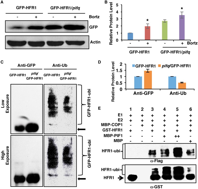
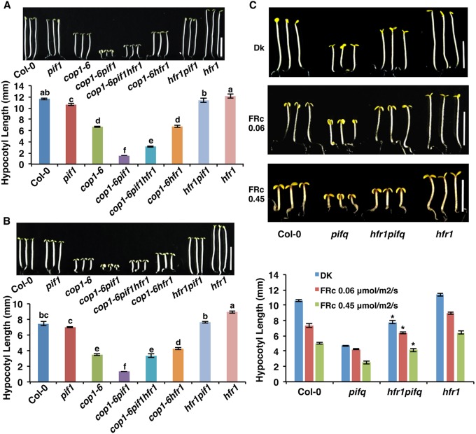
The phytochrome-mediated regulation of photomorphogenesis under red and far-red light conditions involves both positively and negatively acting factors. The positively acting factors (e.g. HY5/HFR1/LAF1 and others) are degraded in the dark to prevent photomorphogenesis. By contrast, the negatively acting factors (e.g. phytochrome-interacting factors or PIFs) are degraded in response to light to promote photomorphogenesis. Here, we show that the negatively acting factor PIF1 is also degraded in the dark by direct heterodimerization with the positively acting factor HFR1. Conversely, PIF1 also promotes the degradation of HFR1 in darkness. PIF1 enhances the poly-ubiquitylation of HFR1 by COP1 in vivo and in vitro In addition, the reciprocal co-degradation of PIF1 and HFR1 is dependent on the 26S proteasome pathway in vivo Genetic evidence shows that the hfr1 mutant partially suppresses the constitutive photomorphogenic phenotypes of cop1-6 pif1 and of the quadruple mutant pifq both in the dark and in far-red light conditions. Taken together, these data uncover a co-degradation mechanism between PIFs and HFR1 that underlies photomorphogenic development in Arabidopsis thaliana.

Keywords: 26S proteasome; E3 ubiquitin ligase; Photomorphogenesis; Reciprocal degradation; bHLH transcription factor.


Conflict of interest statement

Competing interestsThe authors declare no competing or financial interests.

Figures

Fig. 1.
Fig. 1.
PIF1, PIF3, PIF4 and PIF5 are degraded in the dark via the 26S proteasome pathway. (A) Immunoblots showing the level of PIFs in 4-day-old pPIF1:TAP-PIF1, 35S:PIF3-Myc, 35S:PIF4-Myc and 35S:PIF5-Myc dark-grown seedlings. One batch of seedlings was pretreated with 40 µM Bortezomib (Bortz) for 3 h before protein extraction. pif1, pif3, pif4 and pif5 mutants were used as controls. The blot was probed with anti-Myc or anti-RPT5 antibodies. (B) Quantification of TAP-PIF1, PIF3-Myc, PIF4-Myc and PIF5-Myc protein levels using ImageJ. RPT5 was used as a control. The TAP-PIF1, PIF3-Myc, PIF4-Myc and PIF5-Myc protein levels without proteasome inhibitor treatment were set as 100, respectively. Error bars indicate s.d. (n=3).
Fig. 2.
Fig. 2.
HFR1 promotes PIF1 degradation in the dark. (A) Immunoblot shows higher abundance of PIF1 in the hfr1 and cop1-4 hfr1 backgrounds compared with wild-type seedlings. Four-day-old dark-grown seedlings were used for protein extraction. The blot was probed with anti-PIF1 and anti-RPT5 antibodies. (B) Quantification of PIF1 protein level using RPT5 as a control. The letters a-d indicate statistically significant differences between means of protein levels (P<0.05) based on two-way ANOVA analyses. Error bars indicate s.d. (n=7). (C) Immunoblots show the PIF1 (top panel) and GFP-HFR1 (middle panel) and loading control RPT5 (bottom panel) levels in wild-type Col-0, cop1-4, cop1-4 hfr1, cop1-4 hfr1/GFP-HFR1 and cop1-4 hfr1/GFP-HFR1*. Immunoblot was performed as described in A. (D) Quantification of PIF1 protein level using RPT5 as a control. The letters a-d indicate statistically significant differences between means of protein levels (P<0.05) based on two-way ANOVA analyses. Error bars indicate s.d. (n=3).
Fig. 3.
Fig. 3.
HFR1-mediated PIF1 degradation is 26S proteasome dependent. (A) Immunoblot shows the TAP-PIF1 level in cop1-4 and cop1-4 hfr1 background. Total protein was extracted from 4-day-old dark-grown seedlings. One batch of seedlings was pretreated with 40 µM Bortezomib (Bortz) for 3 h before protein extraction. The blot was probed with anti-Myc or anti-RPT5 antibodies. Asterisk indicates a cross-reacting band or proteolytically cleaved product. (B) Quantification of TAP-PIF1 protein level using RPT5 as a control. The letters a-c indicate statistically significant differences among four samples and two treatment conditions (P<0.05) based on two-way ANOVA analyses. Error bars indicate s.d. (n=3). (C) TAP-PIF1 level is higher but the ubiquitylation level is lower in the cop1-4 hfr1 compared with cop1-4 background in darkness. Total protein was extracted from 4-day-old dark-grown seedlings with 40 mM Bortezomib pretreatment for 3 h before protein extraction. TAP-PIF1 was immunoprecipitated using anti-Myc antibody from protein extracts. The immunoprecipitated samples were then separated on 6.5% SDS-PAGE gels and probed with anti-Myc (left) or anti-Ub (right) antibodies. The top and bottom panels are low and high exposures, respectively. Arrow indicates TAP-PIF1. (D) Quantification of TAP-PIF1 and TAP-PIF1-ubi protein levels shown in C. The TAP-PIF1 and TAP-PIF1-ubi protein levels in cop1-4 background were set as 1 respectively. Error bars indicate s.d. (n=3).
Fig. 4.
Fig. 4.
PIFs promote the degradation of HFR1 post-translationally in the dark and under far-red light. (A) Immunoblot shows HFR1 protein level in GFP-HFR1 transgenic plants and in pif1, cop1-6 and cop1-6 pif1 harboring the GFP-HFR1 transgene. Seedlings are grown either in the dark for 4 days or grown in the dark for 21 h and then transferred to FRc (0.45 μmol/m2/s) for 3 days. The blot was probed with anti-GFP or anti-RPT5 antibodies. (B) Bar graph shows GFP-HFR1 protein level in the mutants indicated. For quantification, GFP-HFR1 band intensities were measured from three independent blots using ImageJ, and then normalized against RPT5 levels. GFP-HFR1 dark level was set as 1 and the relative protein levels were calculated. Error bars indicate s.d. Asterisk indicates significant difference (P<0.05) between double and single mutant background. (C) Immunoblot shows HFR1 protein level in the GFP-HFR1 and pifq/GFP-HFR1. RPT5 was used as loading control. Seedlings were grown in the dark or FRc light as described above. (D) Bar graph shows the quantified GFP-HFR1 levels in GFP-HFR1 and pifq/GFP-HFR1. Error bars indicate s.d. Asterisk indicates significant difference between GFP-HFR1 and pifq/GFP-HFR1 in both conditions, respectively (P<0.05). DK, dark.
Fig. 5.
Fig. 5.
PIF1 promotes HFR1 degradation in a ubiquitylation-dependent manner. (A) Immunoblot shows the GFP-HFR1 protein level in GFP-HFR1 and GFP-HFR1/pifq backgrounds. Total protein was extracted from 4-day-old seedlings grown in darkness. One batch of seedlings was pretreated with 40 mM Bortezomib (Bortz) for 3 h before protein extraction. The blot was probed with anti-GFP or anti-actin antibodies. (B) Quantification of GFP-HFR1 protein level using actin as a control. Asterisks indicate statistically significant differences compared with non-Bortezomib treatment for GFP-HFR1 and GFP-HFR1/pifq, respectively (P<0.05). Error bars indicate s.d. (n=4). (C) The protein level of GFP-HFR1 is higher but the ubiquitylation level of GFP-HFR1 is lower in the pifq compared with the GFP-HFR1 background in darkness in vivo. Sample preparation is as described in A. GFP-HFR1 was immunoprecipitated using anti-GFP antibody, and then separated on 8% SDS-PAGE gels and probed with anti-GFP (left) or anti-Ub (right) antibodies. The top and bottom panels are low and high exposures, respectively. The arrow indicates the GFP-HFR1 size. (D) Quantification of GFP-HFR1 and GFP-HFR1-ubi levels for the blot shown in C by ImageJ. The GFP-HFR1 and GFP-HFR1-ubi levels were set as 1, respectively. Error bars indicate s.d. (n=3). (E) PIF1 promotes the ubiquitylation of HFR1 by COP1 in vitro. In vitro ubiquitylation assay was performed using MBP-COP1 as E3 ubiquitin ligase, GST-HFR1 as a substrate, Flag-ubiquitin, UBE1 (E1), UbcH5b (E2) and increasing concentrations of MBP-PIF1. MBP was used as a control. Ubiquitylated GST-HFR1 was detected by anti-Flag antibody (top panel) and anti-GST antibody (bottom panel). The arrow indicates non-ubiquitylated GST-HFR1.
Fig. 6.
Fig. 6.
hfr1 partially suppresses the phenotypes of cop1-6 pif1 and pifq. (A,B) Photographs and bar graphs showing hypocotyl lengths of seedlings of wild type, pif1, cop1-6, cop1-6 pif1, cop1-6 pif1 hfr1, cop1-6 hfr1, hfr1 pif1 and hfr1. Seedlings were grown either in the dark for 5 days (A) or grown in the dark for 21 h then transferred to continuous FRc (0.06 μmol/m2/s) for 4 days (B). Error bars indicate s.d. The letters a-f indicate statistically significant differences between means for hypocotyl lengths (P<0.05) based on two-way ANOVA analyses (n>30, three biological replicates). (C) Photographs and bar graph showing hypocotyl lengths of seedlings of wild type, pifq, hfr1 pifq and hfr1. Seedlings were grown either in the dark for 5 days (top panel) or grown in the dark for 21 h then transferred to FRc for four additional days. Error bars indicate s.d. Asterisks indicate significant difference (P<0.05) compared with pifq (n>30, three biological replicates). Scale bars: 5 mm.

Similar articles

Cited by

References

    1. Bae G. and Choi G. (2008). Decoding of light signals by plant phytochromes and their interacting proteins. Annu. Rev. Plant Biol. 59, 281-311. 10.1146/annurev.arplant.59.032607.092859 - DOI - PubMed
    1. Bauer D., Viczián A., Kircher S., Nobis T., Nitschke R., Kunkel T., Panigrahi K. C. S., Adám, E., Fejes E. Schäfer E. et al. (2004). Constitutive photomorphogenesis 1 and multiple photoreceptors control degradation of phytochrome interacting factor 3, a transcription factor required for light signaling in Arabidopsis. Plant Cell 16, 1433-1445. 10.1105/tpc.021568 - DOI - PMC - PubMed
    1. Bernardo-García S., de Lucas M., Martínez C., Espinosa-Ruiz A., Davière J.-M. and Prat S. (2014). BR-dependent phosphorylation modulates PIF4 transcriptional activity and shapes diurnal hypocotyl growth. Genes Dev. 28, 1681-1694. 10.1101/gad.243675.114 - DOI - PMC - PubMed
    1. Bu Q., Castillon A., Chen F., Zhu L. and Huq E. (2011a). Dimerization and blue light regulation of PIF1 interacting bHLH proteins in Arabidopsis. Plant Mol. Biol. 77, 501-511. 10.1007/s11103-011-9827-4 - DOI - PubMed
    1. Bu Q., Zhu L., Dennis M. D., Yu L., Lu S. X., Person M. D., Tobin E. M., Browning K. S. and Huq E. (2011b). Phosphorylation by CK2 enhances the rapid light-induced degradation of PIF1. J. Biol. Chem. 286, 12066-12074. 10.1074/jbc.M110.186882 - DOI - PMC - PubMed

Publication types

MeSH terms

Substances

LinkOut - more resources