. 2017 May 15;144(10):1807-1819.
doi: 10.1242/dev.141911. Epub 2017 Apr 18.

Excitatory neurons sculpt GABAergic neuronal connectivity in the C. elegans motor circuit

Affiliations

Excitatory neurons sculpt GABAergic neuronal connectivity in the C. elegans motor circuit

Belinda Barbagallo et al. Development. .

Abstract

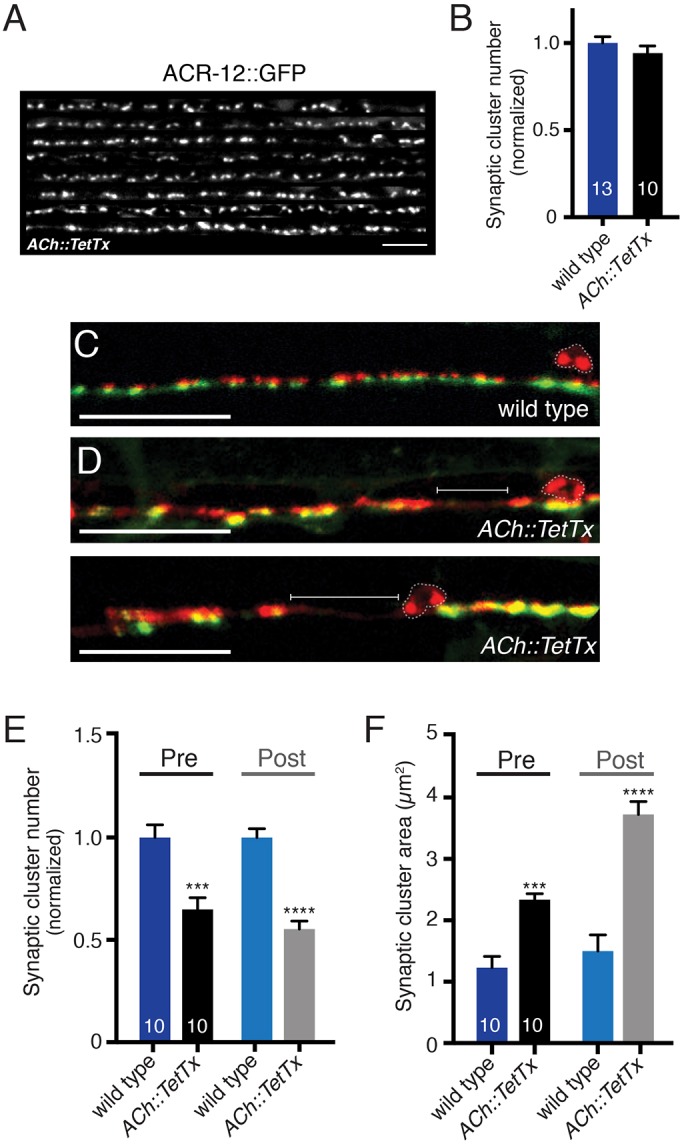
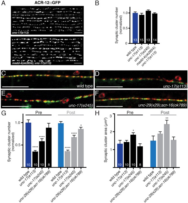
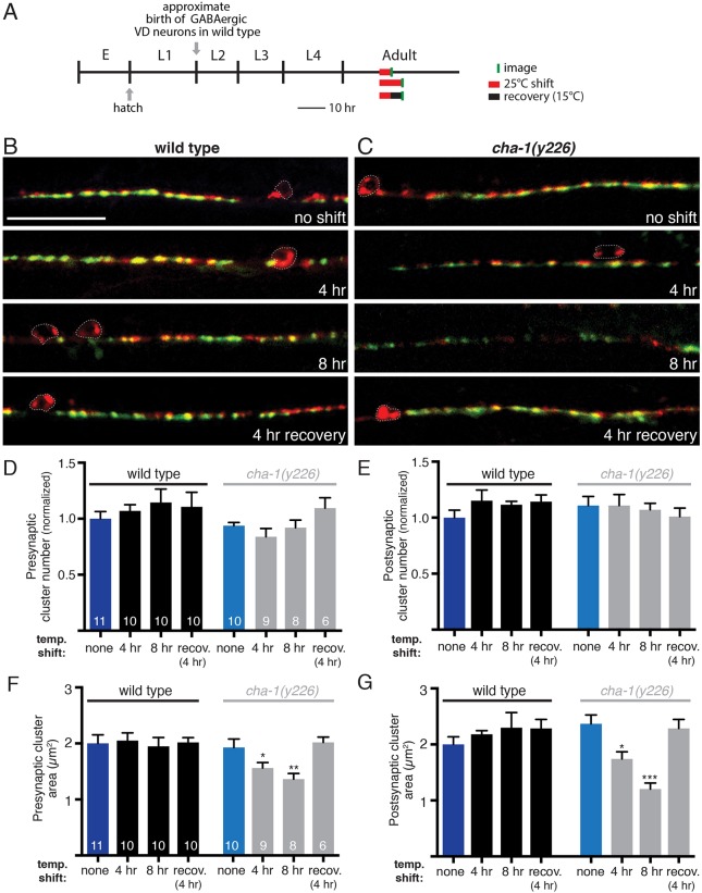
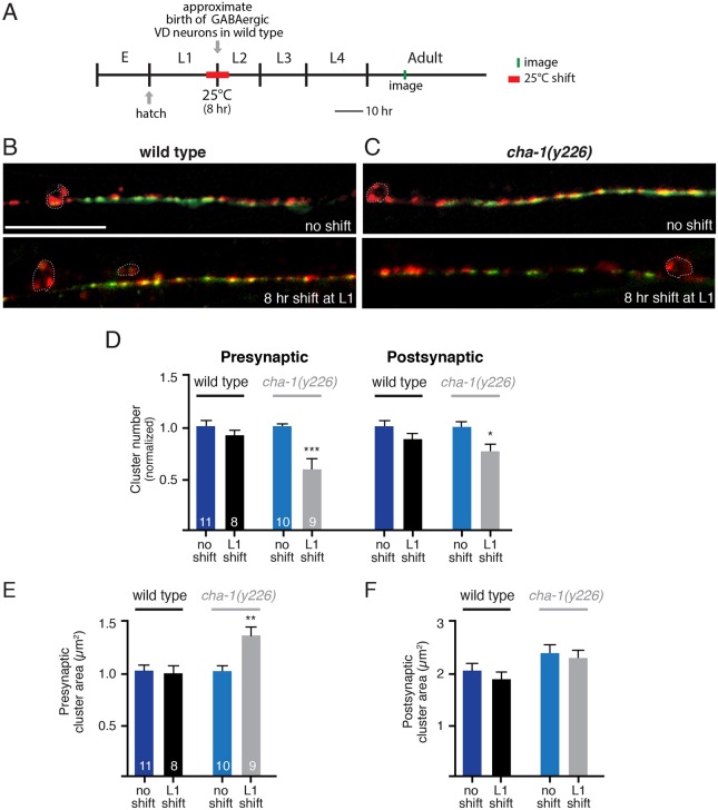
Establishing and maintaining the appropriate number of GABA synapses is key for balancing excitation and inhibition in the nervous system, though we have only a limited understanding of the mechanisms controlling GABA circuit connectivity. Here, we show that disrupting cholinergic innervation of GABAergic neurons in the C. elegans motor circuit alters GABAergic neuron synaptic connectivity. These changes are accompanied by reduced frequency and increased amplitude of GABAergic synaptic events. Acute genetic disruption in early development, during the integration of post-embryonic-born GABAergic neurons into the circuit, produces irreversible effects on GABAergic synaptic connectivity that mimic those produced by chronic manipulations. In contrast, acute genetic disruption of cholinergic signaling in the adult circuit does not reproduce these effects. Our findings reveal that GABAergic signaling is regulated by cholinergic neuronal activity, probably through distinct mechanisms in the developing and mature nervous system.

Keywords: E/I balance; GABA synapse; Neural circuit; Neural development.


Conflict of interest statement

Competing interestsThe authors declare no competing or financial interests.

Figures

Fig. 1.
Fig. 1.
Cholinergic neuromuscular transmission is impaired in unc-3(e151) mutants. (A) The C. elegans motor circuit. Excitatory cholinergic motor neurons (gray) synapse onto inhibitory GABAergic motor neurons (blue) and onto body wall muscles (brown). (B) Representative recordings of endogenous EPSCs from body wall muscles of wild-type and unc-3(e151) mutant animals. Red traces are expanded views of time segments under the red bars in the upper traces. (C) The average frequency of endogenous EPSCs recorded from wild type and unc-3(e151) mutants. Each bar represents the mean±s.e.m., and numbers in bars indicate the n for each genotype in this figure and for all subsequent figures. ***P<0.0001; Student's t-test. (D) Confocal images of one wild-type (top) and two unc-3(e151) (middle and bottom) animals expressing GFP-tagged AChRs in body wall muscles (myo3p::ACR-16::GFP). Scale bar: 5 µm. (E) Normalized receptor cluster number per 75 µm of ventral nerve cord. *P<0.05; Student's t-test.
Fig. 2.
Fig. 2.
unc-3 mutation causes defects in GABAergic transmission and iAChR clustering in GABAergic neurons. (A) Representative recordings of endogenous IPSCs from body wall muscles of wild-type animals and unc-3(e151) mutants. Red traces are expanded views of time segments under the red bars in the upper traces. (B) The average frequency of endogenous IPSCs recorded from wild type and unc-3(e151) mutants. ***P<0.0001, Student's t-test. (C) Cumulative distribution of amplitudes of endogenous IPSCs recorded from wild type and unc-3(e151) mutants. ***P<0.001; Kolmogorov–Smirnov test. (D) Confocal images showing the dorsal nerve cord of eight animals expressing ACR-12::GFP in GABA neurons for the genotypes indicated. Scale bars: 5 µm. (E) Quantification of the average number of receptor clusters per 85 µm of dorsal nerve cord for the genotypes indicated, normalized to wild type. *P<0.05, ****P<0.0001; ANOVA with Dunnett's multiple comparisons test.
Fig. 3.
Fig. 3.
Gross morphological development of GABA motor neurons and muscles does not require cholinergic innervation. (A-C′) Confocal images of the ventral nerve cord and body wall muscles posterior to the vulva of an adult wild-type (A,A′), unc-3(e151) mutant (B,B′) and acr-2(L/S) transgenic (C,C′) animals expressing a membrane-bound GFP in the distal row of body wall muscles (him4p::mCD8::GFP) (A-C) or mCherry in the GABA nervous system (unc-47p::mCherry) (A′-C′). Arrows indicate muscle arm shafts and arrowheads indicate muscle arm termini. Asterisks indicate GABA neuron commissures. Scale bars: 10 µm (A-C); 20 µm (A′-C′). (D) Quantification of the percentage of the ventral nerve cord region covered by muscle membrane. ns, not significant; ANOVA with Dunnett's multiple comparisons test.
Fig. 4.
Fig. 4.
Impaired cholinergic innervation alters the distribution and size of GABA synapses. (A) Diagram depicting the location of fluorescent reporters used to label GABA pre- (unc-47p::mCherry::RAB-3) and postsynaptic (UNC-49::GFP) structures. (B-F) Confocal images of the ventral nerve cord in adult wild type (B), unc-3(n3435) mutants (C), unc-3(e151) mutants (D), unc-3(e151) mutants expressing an unc-3 cDNA rescuing array (unc-3p::unc-3 cDNA) (E), and transgenic acr-2(L/S) animals (F) co-expressing unc-47p::mCherry::RAB-3 in GABA motor neurons with UNC-49::GFP in body wall muscles. Note areas devoid of synaptic clusters (brackets) and enlarged clusters in unc-3 mutants and acr-2(L/S) transgenic animals. Motor neuron cell bodies are outlined in this figure and all subsequent figures. Scale bars: 20 µm. (G) Average number of pre- and postsynaptic clusters per 50 µm, normalized to wild type. (H) Average size of pre- and postsynaptic clusters at GABA synapses. *P<0.05, **P<0.01, ****P<0.0001 compared with either control or unc-3 mutant as indicated; ANOVA with Dunnett's multiple comparisons test. (I,J) Confocal images of the ventral cord in wild type or unc-3(e151) mutants expressing the GFP-tagged synaptic vesicle marker synaptobrevin (SNB-1::GFP) (I), or the Rim1 homolog (UNC-10::mCherry) (J). Scale bars: 20 µm. (K) Quantification of synaptic cluster number per 50 μm for each marker, normalized to wild type. (L) Quantification of presynaptic cluster area for genotypes indicated. **P<0.01, ***P<0.0001; Student's t-test.
Fig. 5.
Fig. 5.
Vesicular release from cholinergic motor neurons shapes GABA synaptic outputs. (A) Confocal images showing the dorsal nerve cord of eight animals co-expressing ACR-12::GFP in GABA neurons and tetanus toxin in cholinergic neurons. Scale bar: 5 µm. (B) Quantification of the average number of receptor clusters per 85 µm of dorsal nerve cord for the genotypes indicated, normalized to wild type. (C,D) Merged confocal images of unc-47p::mCherry::RAB-3 and UNC-49::GFP labeling in the ventral nerve cord of adult wild-type (C) and transgenic animals with specific TetTx expression in cholinergic neurons (D). Brackets indicate areas devoid of synaptic clusters. Scale bars: 20 µm. (E) Average number of pre- and postsynaptic clusters per 50 µm for each genotype, normalized to wild type. (F) Average area of pre- and postsynaptic clusters at GABA synapses for each genotype as indicated. ***P<0.001, ****P<0.0001; Student's t-test.
Fig. 6.
Fig. 6.
Reduced cholinergic transmission alters GABA synapse density and localization. (A) Confocal images showing the dorsal nerve cord of eight unc-17(e113) and madd-4(ok2854);unc-17(e113) animals expressing ACR-12::GFP in GABA neurons. Scale bars: 5 µm. (B) Quantification of the average number of receptor clusters per 85 µm of dorsal nerve cord for the genotypes indicated, normalized to wild type. (C-F) Merged confocal images of unc-47p::mCherry::RAB-3 and UNC-49::GFP labeling in the ventral nerve cord region of adult wild-type animals (C), unc-17(e113)/vAChT mutants (D), unc-17(e245)/vAChT mutants (E) or unc-29(x29);acr-16(ok789) double mutants, which lack functional muscle iAChRs (F). Scale bars: 20 µm. (G) Average number of GABA pre- and postsynaptic clusters per 50 µm for each genotype, normalized to wild type. (H) Average size of GABA pre- and postsynaptic clusters. *P<0.05, **P<0.01, ****P<0.0001; ANOVA with Dunnett's multiple comparisons test.
Fig. 7.
Fig. 7.
Acute reduction of cholinergic transmission during adulthood produces reversible decreases in GABA synapse size. (A) Diagram indicating predicted timeline of wild-type C. elegans development at 15°C and temperature shifts employed. Approximate time of hatch is indicated. Temperature shifts to 25°C (red) and time of imaging (green) are indicated. E, embryo. (B,C) Merged confocal images of unc-47p::mCherry::RAB-3 and UNC-49::GFP labeling in the ventral nerve cord region of adult wild-type animals (B) or cha-1(y226) mutants (C) following a shift to 25°C for the durations indicated. Scale bar: 20 µm. (D,E) Average number of GABA presynaptic (D) and postsynaptic (E) clusters per 50 µm for wild type (black) and cha-1(y226) mutants (gray). (F,G) Average size of GABA presynaptic (F) and postsynaptic (G) clusters for wild type (black) and cha-1(y226) mutants (gray). *P<0.05, **P<0.01, ***P<0.0001; ANOVA with Dunnett's multiple comparisons test.
Fig. 8.
Fig. 8.
Reduced cholinergic neurotransmission during a period of GABAergic synaptogenesis alters GABAergic synaptic connectivity. (A) Diagram indicating predicted timeline of wild-type C. elegans development at 15°C and temperature shifts employed. Approximate time of hatch is indicated. (B,C) Confocal images of the ventral nerve cord in adult wild-type (B) and cha-1(y226) (C) temperature-sensitive mutant animals co-expressing GABA pre- and postsynaptic markers (unc-47p::mCherry::RAB-3 and UNC-49::GFP) grown at the permissive temperature and after an 8 h shift to the non-permissive temperature during the first larval stage. Note that the representative image for the cha-1 no shift control group shown in C is the same as that displayed in Fig. 7C. Scale bar: 20 µm. (D) Average number of GABA presynaptic and postsynaptic clusters per 50 µm for wild type (black) and cha-1(y226) mutants (gray). (E,F) Average size of GABA presynaptic (E) and postsynaptic (F) clusters for wild type (black) and cha-1(y226) mutants (gray). *P<0.05, **P<0.01, ***P<0.0001; Student's t-test.

Similar articles

Cited by

References

    1. Ackley B. D., Harrington R. J., Hudson M. L., Williams L., Kenyon C. J., Chisholm A. D. and Jin Y. (2005). The two isoforms of the Caenorhabditis elegans leukocyte-common antigen related receptor tyrosine phosphatase PTP-3 function independently in axon guidance and synapse formation. J. Neurosci. 25, 7517-7528. 10.1523/JNEUROSCI.2010-05.2005 - DOI - PMC - PubMed
    1. Alfonso A., Grundahl K., Duerr J. S., Han H. P. and Rand J. B. (1993). The Caenorhabditis elegans unc-17 gene: a putative vesicular acetylcholine transporter. Science 261, 617-619. 10.1126/science.8342028 - DOI - PubMed
    1. Bamber B. A., Beg A. A., Twyman R. E. and Jorgensen E. M. (1999). The Caenorhabditis elegans unc-49 locus encodes multiple subunits of a heteromultimeric GABA receptor. J. Neurosci. 19, 5348-5359. - PMC - PubMed
    1. Barbagallo B., Prescott H. A., Boyle P., Climer J. and Francis M. M. (2010). A dominant mutation in a neuronal acetylcholine receptor subunit leads to motor neuron degeneration in Caenorhabditis elegans. J. Neurosci. 30, 13932-13942. 10.1523/JNEUROSCI.1515-10.2010 - DOI - PMC - PubMed
    1. Bhattacharya R., Touroutine D., Barbagallo B., Climer J. Lambert C. M., Clark C. M., Alkema M. J. and Francis M. M. (2014). A conserved dopamine-cholecystokinin signaling pathway shapes context-dependent Caenorhabditis elegans behavior. PLoS Genet. 10, e1004584 https://doi.org/10.1371/journal.pgen.1004584 - DOI - PMC - PubMed

Publication types

MeSH terms

LinkOut - more resources