. 2017 Jun 9;292(23):9652-9665.
doi: 10.1074/jbc.M117.786855. Epub 2017 Apr 18.

Structure and characterization of a class 3B proline utilization A: Ligand-induced dimerization and importance of the C-terminal domain for catalysis

Affiliations

Structure and characterization of a class 3B proline utilization A: Ligand-induced dimerization and importance of the C-terminal domain for catalysis

David A Korasick et al. J Biol Chem. .

Abstract

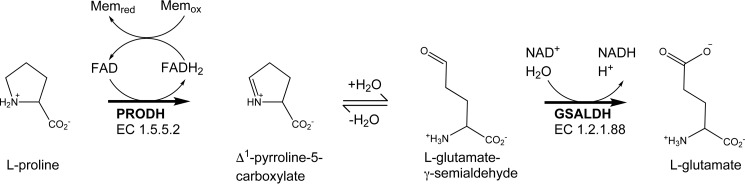
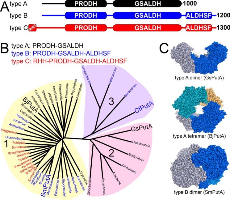
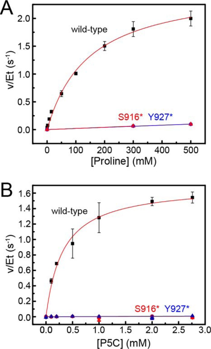
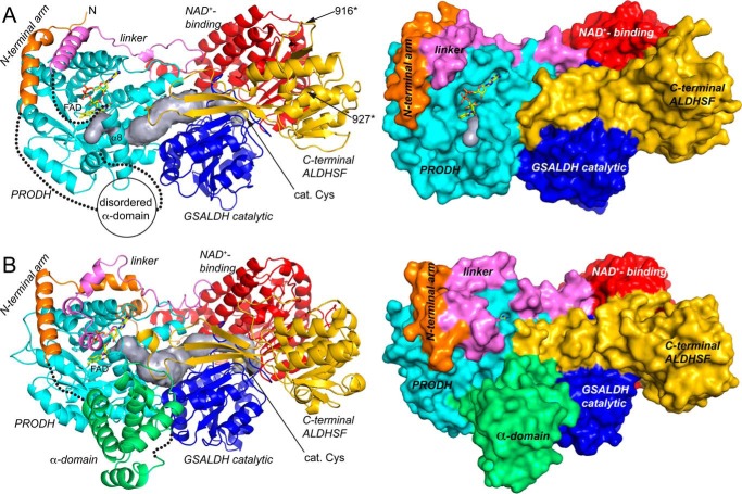
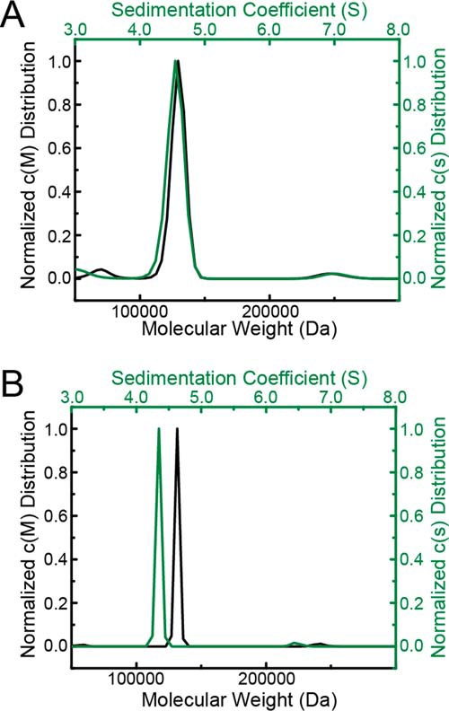
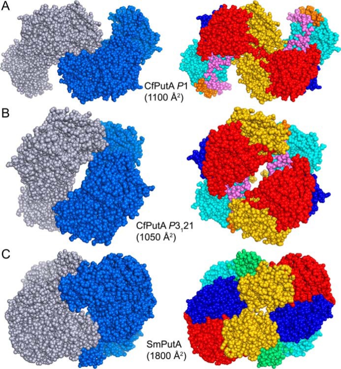
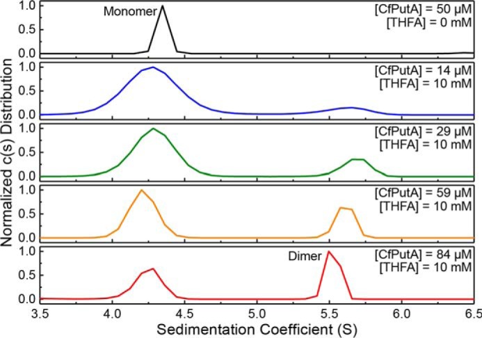
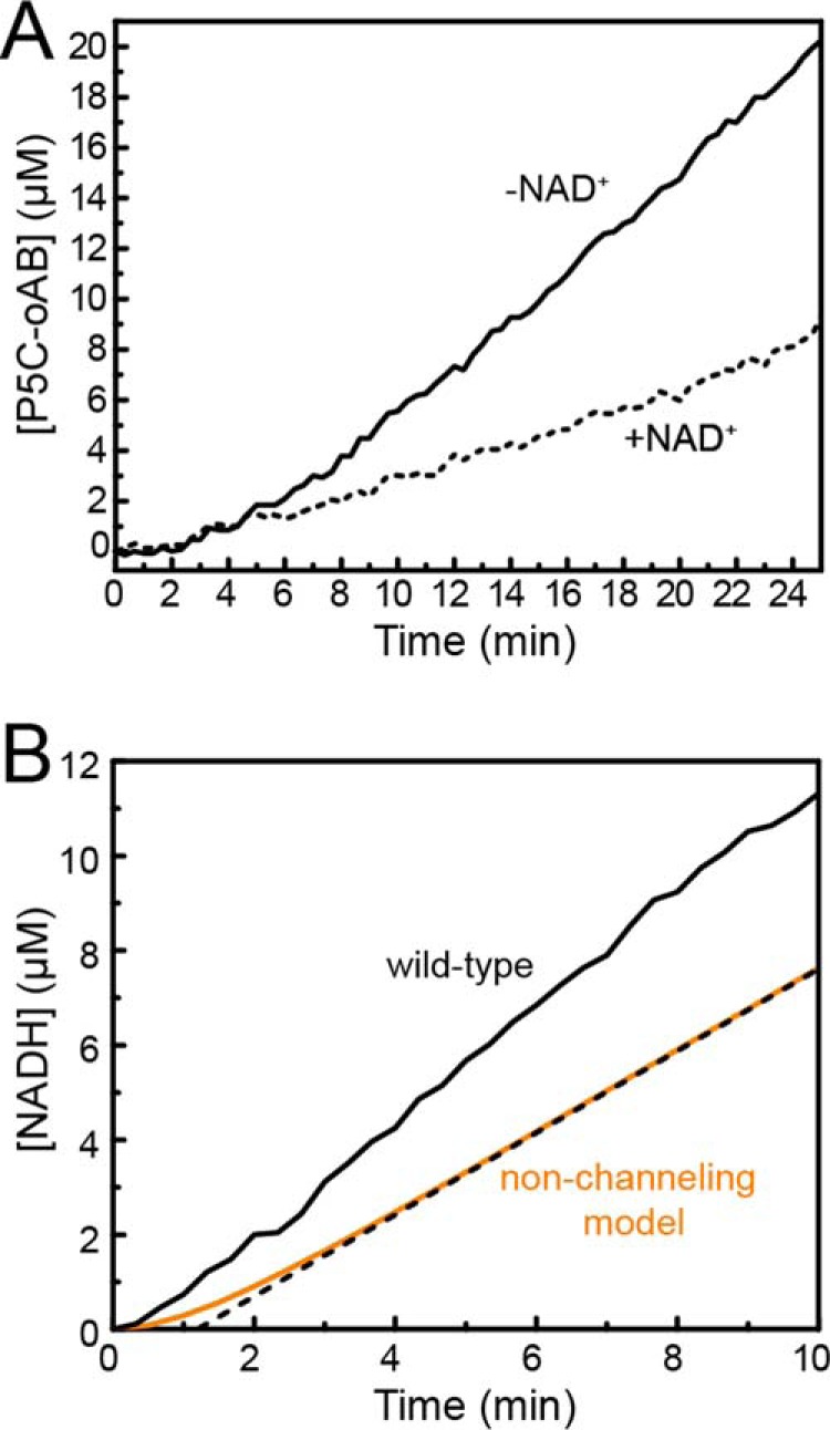
The bifunctional flavoenzyme proline utilization A (PutA) catalyzes the two-step oxidation of proline to glutamate using separate proline dehydrogenase (PRODH) and l-glutamate-γ-semialdehyde dehydrogenase active sites. Because PutAs catalyze sequential reactions, they are good systems for studying how metabolic enzymes communicate via substrate channeling. Although mechanistically similar, PutAs vary widely in domain architecture, oligomeric state, and quaternary structure, and these variations represent different structural solutions to the problem of sequestering a reactive metabolite. Here, we studied PutA from Corynebacterium freiburgense (CfPutA), which belongs to the uncharacterized 3B class of PutAs. A 2.7 Å resolution crystal structure showed the canonical arrangement of PRODH, l-glutamate-γ-semialdehyde dehydrogenase, and C-terminal domains, including an extended interdomain tunnel associated with substrate channeling. The structure unexpectedly revealed a novel open conformation of the PRODH active site, which is interpreted to represent the non-activated conformation, an elusive form of PutA that exhibits suboptimal channeling. Nevertheless, CfPutA exhibited normal substrate-channeling activity, indicating that it isomerizes into the active state under assay conditions. Sedimentation-velocity experiments provided insight into the isomerization process, showing that CfPutA dimerizes in the presence of a proline analog and NAD+ These results are consistent with the morpheein model of enzyme hysteresis, in which substrate binding induces conformational changes that promote assembly of a high-activity oligomer. Finally, we used domain deletion analysis to investigate the function of the C-terminal domain. Although this domain contains neither catalytic residues nor substrate sites, its removal impaired both catalytic activities, suggesting that it may be essential for active-site integrity.

Keywords: X-ray crystallography; analytical ultracentrifugation; bifunctional enzyme; dehydrogenase; enzyme kinetics; flavoprotein; oligomerization; substrate channeling.


Conflict of interest statement

The authors declare that they have no conflicts of interest with the contents of this article

Figures

Figure 1.
Figure 1.
The reactions of proline catabolism.
Figure 2.
Figure 2.
Classification and diversity of PutAs. A, the three domain architectures of PutAs. B, phylogenetic tree based on global sequence alignments of PutAs. PutAs with architecture types A, B, and C are indicated by black, blue, and red type, respectively. The four PutAs for which crystal structures have been obtained are listed in large type. The alignments were calculated with Clustal Omega (56) and visualized with DrawTree (57). C, structures of PutA oligomers that have been validated by crystallography and SAXS (PDB codes 4NMA, 3HAZ, and 5KF6).
Figure 3.
Figure 3.
Steady-state kinetics data. A, dependence of PRODH activity on proline concentration. B, dependence of GSALDH activity on P5C concentration. For both panels, results are shown for wild-type CfPutA (black squares), CfPutA S916* (red triangles), and CfPutA Y927* (blue triangles). Fits to the Michaelis-Menten equation for wild-type CfPutA are represented by the red curves. Linear regression was used for the mutant variants to guide the eye and to estimate kcat/Km for PRODH activity. Analysis was done using Origin 2016.
Figure 4.
Figure 4.
Structure of the CfPutA monomer and comparison to SmPutA. A, backbone and surface representations of CfPutA. The domains have different colors, with the N-terminal arm in orange, PRODH barrel in cyan, PRODH-GSALDH linker in violet, GSALDH NAD+-binding in red, GSALDH catalytic in blue, and C-terminal ALDHSF in gold. The silver surface represents the substrate-channeling tunnel. The FAD is shown in yellow sticks. The dotted lines indicate disordered residues 33–118 (α-domain) and 421–434 (which connect α8 to the PRODH-GSALDH linker). The sites of domain deletion mutations are indicated as 916* and 927*. B, backbone and surface representations of SmPutA. The domains are colored as in A. The α-domain, which is disordered in CfPutA, is colored green.
Figure 5.
Figure 5.
Comparison of the C-terminal ALDHSF domains of CfPutA and SmPutA. A, the ALDHSF domain of CfPutA. The truncation points for the S916* and Y927* C-terminal deletion mutants are marked by arrowheads. B, the ALDHSF domain of SmPutA. In both panels, the diagram shows the topology of the Rossmann fold part of the ALDHSF domain.
Figure 6.
Figure 6.
Unexpected FAD conformation. A, 2.7 Å resolution electron density for the FAD in CfPutA. The cage represents a simulated annealing σA-weighted FoFc omit map (3.0σ). The inset shows an edge-on view of the isoalloxazine ring. B, superposition of the FAD in CfPutA (yellow) and the 2-electron-reduced FAD in GsPutA (gray). The inset shows an edge-on view of the isoalloxazine ring. Note that the two FADs have the same ribityl conformation but different butterfly bend angles. C, a typical oxidized PutA FAD (from GsPutA). Note that the ribityl conformation differs from that in B.
Figure 7.
Figure 7.
Sedimentation-velocity analysis of CfPutA in the absence of active-site ligands. A, sedimentation coefficient and molecular mass distributions for CfPutA at 0.8 mg/ml (7 μm). B, sedimentation coefficient and molecular mass distributions for CfPutA at 6 mg/ml (50 μm).
Figure 8.
Figure 8.
Dimeric assemblies of CfPutA and SmPutA. A, the largest protein-protein interface in the CfPutA P1 crystal form. B, the largest protein-protein interface in the CfPutA trigonal crystal form. C, the bona fide dimer of SmPutA, which has been validated by SAXS and crystallography (PDB code 5KF6). The interfacial surface areas are listed for each assembly. On the left side, the chains have different colors. On the right side, the domains are colored as in Fig. 4: N-terminal arm (orange), α-domain (green), PRODH barrel (cyan), PRODH-GSALDH linker (violet), NAD+-binding (red), GSALDH catalytic (blue), and C-terminal ALDHSF (gold).
Figure 9.
Figure 9.
CfPutA undergoes concentration-dependent oligomerization in the presence of active-site ligands. Sedimentation-velocity experiments were performed at the indicated CfPutA concentrations in the absence (top panel) or presence (bottom four panels) of the proline analog THFA (10 mm) and NAD+ (1 mm).
Figure 10.
Figure 10.
Evidence for substrate channeling in CfPutA. A, P5C-oAB-trapping experiments performed in the absence (solid lines) or presence (dashed lines) of NAD+. B, transient-time analysis of CfPutA. Experimental data from a PRODH-GSALDH-coupled assay are represented in solid black. A non-channeling model calculated from Equation 1 is represented in orange. The dotted black line represents extrapolation of the predicted steady-state rate to estimate a transient time of 1.2 min.
Figure 11.
Figure 11.
Deletion of the ALDHSF domain does not result in apparent destabilization of CfPutA tertiary structure. Thermal shift assay melt curves are shown for wild-type CfPutA (black), CfPutA S916* (red), and CfPutA Y927* (blue). The horizontal black dashed line represents 0.5 of the total fraction unfolded. The three vertical dashed lines indicate the T0.5 of each protein. The plotted curves are the average of samples measured in triplicate normalized to fraction unfolded.

Similar articles

Cited by

References

    1. Tanner J. J., and Becker D. F. (2013) PutA and proline metabolism. in Handbook of Flavoproteins, Vol. 1 (Hille R., Miller S. M., and Palfey B. A., eds) pp. 31–56, De Gruyter, Berlin
    1. Srivastava D., Schuermann J. P., White T. A., Krishnan N., Sanyal N., Hura G. L., Tan A., Henzl M. T., Becker D. F., and Tanner J. J. (2010) Crystal structure of the bifunctional proline utilization A flavoenzyme from Bradyrhizobium japonicum. Proc. Natl. Acad. Sci. U.S.A. 107, 2878–2883 - PMC - PubMed
    1. Arentson B. W., Luo M., Pemberton T. A., Tanner J. J., and Becker D. F. (2014) Kinetic and structural characterization of tunnel-perturbing mutants in Bradyrhizobium japonicum proline utilization A. Biochemistry 53, 5150–5161 - PMC - PubMed
    1. Luo M., Christgen S., Sanyal N., Arentson B. W., Becker D. F., and Tanner J. J. (2014) Evidence that the C-terminal domain of a type B PutA protein contributes to aldehyde dehydrogenase activity and substrate channeling. Biochemistry 53, 5661–5673 - PMC - PubMed
    1. Moxley M. A., Sanyal N., Krishnan N., Tanner J. J., and Becker D. F. (2014) Evidence for hysteretic substrate channeling in the proline dehydrogenase and Δ1-pyrroline-5-carboxylate dehydrogenase coupled reaction of proline utilization A (PutA). J. Biol. Chem. 289, 3639–3651 - PMC - PubMed

Publication types

MeSH terms

LinkOut - more resources