Review
. 2017 Jun;15(3):135-141.
doi: 10.1007/s11914-017-0359-y.

Parathyroid Diseases and T Cells

Affiliations
Review

Parathyroid Diseases and T Cells

M Neale Weitzmann et al. Curr Osteoporos Rep. 2017 Jun.

Abstract

Purpose of review: This review summarizes studies into the permissive role of T cells in the bone catabolic effects of hyperparathyroidism and parathyroid hormone (PTH).

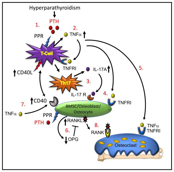
Recent findings: Work in animals combined with recent translational studies in humans now highlight the potent amplificatory action of T cells on PTH-induced bone resorption. Mechanistic animal studies reveal a complex pathway by which PTH exploits natural self-renewal functions of CD4+ T cells, to drive TNFα production that promotes formation of IL-17A secreting Th17 T cells. TNFα and IL-17 further amplify osteoblastic receptor activator of NF-κB ligand (RANKL) production and down-modulate osteoprotegerin (OPG), establishing conditions propitious for osteoclastic bone resorption. These findings are consistent with, and add to, the traditional view of PTH-induced bone loss involving only osteoblast-lineage cells. T cells potently amplify traditional pathways and provide permissive costimulatory signals to bone marrow stromal cells, facilitating the development of an increased RANKL/OPG ratio favourable to bone resorption and bone loss.

Keywords: Hyperparathyroidism; Osteoimmunology; Osteoporosis; PTH; Parathyroid hormone; T cells.


Figures

Figure 1
Figure 1. Model of T cell dependent mechanism of cPTH-induced bone loss
(1.) Binding of PTH to its receptor (PPR) on the CD4+ T cell initiates (2.) production of TNFα, a cytokine that binds to a Type I receptor (TNFRI) and promotes differentiation of the T cell into (3.) a Th17 helper cell secreting IL-17A. (4.) Both of TNFα and IL-17A bind to receptors (TNFRI and IL-17R respectively) on BMSC, osteoblasts and osteocytes. (5.) TNFα further binds to osteoclastic cells amplifying RANKL activity on the differentiating osteoclast and stimulating bone resorption. (6.) TNFα and IL-17A promote production of RANKL and suppress production of OPG, the RANKL inhibitor, creating a balance of RANKL to OPG favourable to increased osteoclastic differentiation. (7.) TNFα further upregulates expression of CD40 on BMSC allowing a costimulatory signal to be transduced from the T cells though expression of CD40L. This signal primes the osteoblast to further respond to direct PTH stimulation to ensure robust RANKL production. (8) RANKL binds to its receptor RANK on osteoclast lineage cells causing osteoclast precursor differentiation and fusion into mature osteoclasts that resorb bone leading to bone loss. Model adapted from Li et al., [39].

References

    1. Marcocci C, Cetani F. Clinical practice. Primary hyperparathyroidism. N Engl J Med. 2011;365(25):2389–97. - PubMed
    1. Holick MF. Vitamin D deficiency. N Engl J Med. 2007;357(3):266–81. - PubMed
    1. Pacifici R. T cells: critical bone regulators in health and disease. Bone. 2010;47(3):461–71. - PMC - PubMed
    1. Calvi LM, Sims NA, Hunzelman JL, Knight MC, Giovannetti A, Saxton JM, et al. Activated parathyroid hormone/parathyroid hormone-related protein receptor in osteoblastic cells differentially affects cortical and trabecular bone. J Clin Invest. 2001;107(3):277–86. - PMC - PubMed
    1. Ben-awadh AN, Delgado-Calle J, Tu X, Kuhlenschmidt K, Allen MR, Plotkin LI, et al. Parathyroid hormone receptor signaling induces bone resorption in the adult skeleton by directly regulating the RANKL gene in osteocytes. Endocrinology. 2014;155(8):2797–809. - PMC - PubMed

MeSH terms

LinkOut - more resources