. 2017 Jul 15;595(14):4803-4821.
doi: 10.1113/JP273872. Epub 2017 May 25.

Role of mucosa in generating spontaneous activity in the guinea pig seminal vesicle

Affiliations

Role of mucosa in generating spontaneous activity in the guinea pig seminal vesicle

Mitsue Takeya et al. J Physiol. .

Abstract

Key points: The mucosa may have neuron-like functions as urinary bladder mucosa releases bioactive substances that modulate sensory nerve activity as well as detrusor muscle contractility. However, such mucosal function in other visceral organs remains to be established. The role of mucosa in generating spontaneous contractions in seminal vesicles (SVs), a paired organ in the male reproductive tract, was investigated. The intact mucosa is essential for the generation of spontaneous phasic contractions of SV smooth muscle arising from electrical slow waves and corresponding increases in intracellular Ca2+ . These spontaneous events primarily depend on Ca2+ handling by sarco-endoplasmic reticulum Ca2+ stores. A population of mucosal cells developed spontaneous rises in intracellular Ca2+ relying on sarco-endoplasmic reticulum Ca2+ handling. The spontaneously active cells in the SV mucosa appear to drive spontaneous activity in smooth muscle either by sending depolarizing signals and/or by releasing humoral substances.

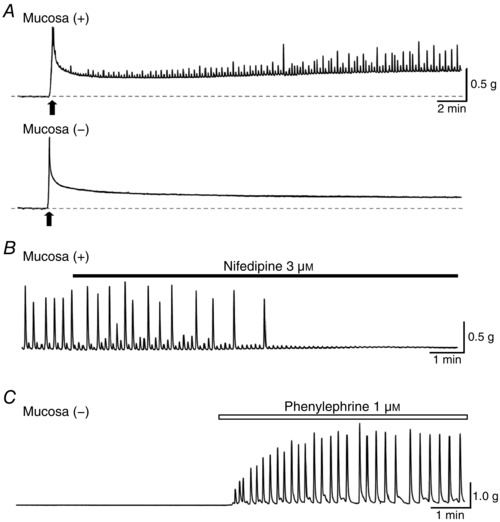
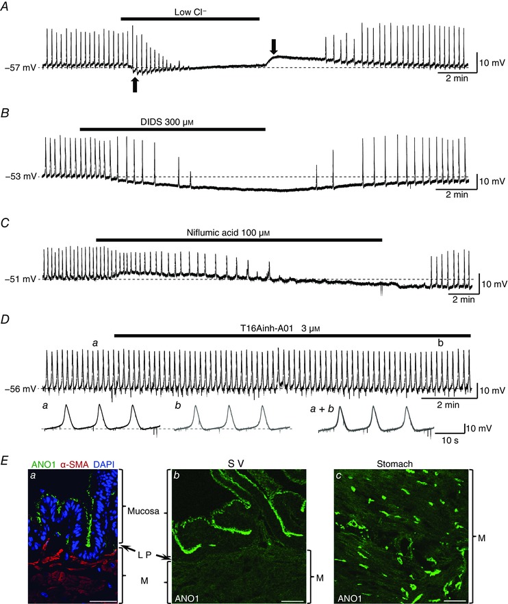
Abstract: The role of the mucosa in generating the spontaneous activity of guinea-pig seminal vesicle (SV) was explored. Changes in contractility, membrane potential and intracellular Ca2+ dynamics of SV smooth muscle cells (SMCs) were recorded using isometric tension recording, intracellular microelectrode recording and epi-fluorescence Ca2+ imaging, respectively. Mucosa-intact but not mucosa-denuded SV preparations generated TTX- (1 μm) resistant spontaneous phasic contractions that were abolished by nifedipine (3 μm). Consistently, SMCs developed mucosa-dependent slow waves (SWs) that triggered action potentials and corresponding Ca2+ flashes. Nifedipine (10 μm) abolished the action potentials and spontaneous contractions, while suppressing the SWs and Ca2+ flashes. Both the residual SWs and spontaneous Ca2+ transients were abolished by cyclopiazonic acid (CPA, 10 μm), a sarco-endoplasmic reticulum Ca2+ -ATPase (SERCA) inhibitor. DIDS (300 μm) and niflumic acid (100 μm), blockers for Ca2+ -activated Cl- channels (CACCs), or low Cl- solution also slowed or prevented the generation of SWs. In SV mucosal preparations detached from the muscle layer, a population of mucosal cells generated spontaneous Ca2+ transients that were blocked by CPA but not nifedipine. These results suggested that spontaneous contractions and corresponding Ca2+ flashes in SV SMCs arise from action potential generation due to the opening of L-type voltage-dependent Ca2+ channels. Spontaneous Ca2+ transients appear to primarily result from Ca2+ release from sarco-endoplasmic reticulum Ca2+ stores to activate CACCs to develop SWs. The mucosal cells firing spontaneous Ca2+ transients may play a critical role in driving spontaneous activity of SV smooth muscle either by sending depolarizing signals or by releasing humoral substances.

Keywords: intracellular Ca2+ release; mucosa; seminal vesicle; slow wave; spontaneous contraction.


Figures

Figure 1
Figure 1. Morphological properties of mucosa‐intact and mucosa‐denuded SV smooth muscle
A, scanning electron micrographs of the mucosal surface of the mucosa‐intact [Mucosa (+)] and mucosa‐denuded [Mucosa (−)] reversed ring SV preparations. Folding of the intact mucosa is evident in mucosa (+) but not mucosa (−) preparations. B, light micrographs of haematoxylin‐eosin‐stained coronal sections of reversed ring preparations of SV. Mucosa (+) preparation consists of inner mucosa, thin lamina propria (LP) and external muscular layer (M). In the mucosa (−) preparation, only loose lamina propria and muscular layer were left. In both mucosa (+) and mucosa (−) preparations, circular muscle layer is obvious just beneath the lamina propria.
Figure 2
Figure 2. Mucosa dependence of spontaneous contractions in SV
A, spontaneous phasic contractions (upward deflections) were periodically developed in mucosa‐intact [Mucosa (+)] but not mucosa‐denuded [Mucosa (−)] SV preparations. Black arrows indicate the timing of initial stretching to 1 g. Dotted base lines indicate 0 g. B, nifedipine (3 μm) abolished the spontaneous contractions in the mucosa‐intact SV. C, in mucosa‐denuded SV that did not generate spontaneous contractions, bath‐applied phenylephrine (1 μm) induced large oscillatory contractions.
Figure 3
Figure 3. Spontaneous and nerve‐evoked contractions in SV
A, in mucosa‐intact SV that exhibited spontaneous contractions, electrical field stimulation (EFS) triggered TTX‐ (1 μm) sensitive, nerve‐evoked contractions (asterisks). Lower traces show expanded recordings in control (a) and in TTX (1 μm, b). Traces a and b were obtained at the timings indicated by the corresponding characters in the upper trace. B, in mucosa‐denuded SV that did not generate spontaneous contractions, EFS triggered nerve‐evoked contractions (asterisks) that were reversibly blocked by TTX (0.1 μm). Trains of EFS (5 V, 0.1 ms, 30 Hz, 3 s) indicated by black bars were applied at 4 min intervals in A and B.
Figure 4
Figure 4. Mucosa dependence of slow waves and action potentials in SV smooth muscle
A, light micrographs of haematoxylin‐eosin‐stained coronal sections of the inner circular smooth muscle preparations of SV with and without intact mucosa similar to those used for recordings of membrane potentials or intracellular Ca2+ dynamics. In the right photograph of the mucosa‐denuded preparation [Mucosa (−)], some mucosa was left attached to indicate the mucosal side (arrow). B, in a mucosa‐intact preparation, SV smooth muscle developed spontaneous slow waves (SWs) with superimposed action potentials. Lower trace shows an SW with an expanded time scale to show measured parameters: RMP, resting membrane potential; AMP (SW), peak amplitude of SW; HW, half‐width of SW. C, in a mucosa‐denuded preparation, SV smooth muscle did not generate SWs nor spontaneous action potentials. D, in a ‘quiescent’ mucosa‐denuded SV smooth muscle, depolarizing current injection (200 pA for 400 ms) evoked several action potentials.
Figure 5
Figure 5. Effects of nifedipine on action potentials and SWs in mucosa‐intact SV smooth muscle
A, in a mucosa‐intact SV preparation, nifedipine (1 μm) abolished superimposed action potentials, leaving SWs. Lower traces show SWs in control (a) and in nifedipine (1 μm, b) with an expanded time scale. Traces a and b were obtained at the timings indicated by the corresponding characters in the upper trace. B, in another mucosa‐intact SV preparation, nifedipine (10 μm) abolished superimposed action potentials, reduced the amplitude of SWs and also depolarized the membrane by 10 mV. Lower traces show SWs in control (a) and in nifedipine (10 μm, b) with an expanded time scale. Traces a and b were obtained at the timings indicated by the corresponding characters in the upper trace. Dotted line indicates RMP in control. Summarized data of the effects of nifedipine (1 and 10 μm) on the peak amplitude [AMP (SW), C] and the frequency (D) of the SWs. The closed circles connected by a line indicate data obtained from identical cells.
Figure 6
Figure 6. Mucosa dependence of spontaneous Ca2+activity in SV smooth muscle
A, sequential Cal‐520 fluorescence images (frame interval;107 ms) demonstrate spontaneous Ca2+ flashes in the mucosa‐intact SV. Scale bars = 30 μm. Spontaneous Ca2+ flashes were generated almost synchronously across SV smooth muscle cells. Middle traces show that Ca2+ flashes recorded from three regions of interest (ROIs: indicated by circles in upper right panel) were generated synchronously. Nifedipine (10 μm) greatly suppressed spontaneous Ca2+ flashes. Lower traces were obtained in control (a) and in nifedipine (10 μm, b) with an expanded time scale. Traces a and b were obtained at the timings indicated by the corresponding characters in the middle trace. The numbers of the traces correspond to the number of ROIs in the upper panel. Note that synchrony was preserved for the nifedipine‐resistant spontaneous Ca2+ transients. Effects of nifedipine (10 μm) on the peak amplitude (B) and frequency (C) of spontaneous Ca2+ flashes in mucosa‐intact SVs were summarized (n = 5). D, in a mucosa‐denuded SV that did not generate spontaneous Ca2+ transients, phenylephrine (1 μm) evoked synchronous oscillatory Ca2+ transients.
Figure 7
Figure 7. Blockade of SERCA abolished nifedipine‐resistant SWs and spontaneous Ca2+ transients in mucosa‐intact SV smooth muscle
A, in a nifedipine‐ (10 μm) pretreated preparation that exhibited SWs, CPA (10 μm) depolarized the membrane by about 5 mV and prevented the generation of SWs. B, in another nifedipine‐ (10 μm) pretreated preparation that developed spontaneous Ca2+ transients, CPA (10 μm) increased the basal Ca2+ level and abolished Ca2+ transients. Dotted line in A indicates RMP.
Figure 8
Figure 8. Role of CACCs in the generation of SWs in mucosa‐intact SV
A, in a nifedipine‐ (10 μm) pretreated preparation that exhibited SWs, lowering [Cl]o prevented the generation of SWs. Negative and positive shifts of the baseline upon the reduction and restoration of [Cl]o, respectively (black arrows), were due to liquid junction potential. Subtraction of the measured liquid junction potential revealed that low Cl solution depolarized the membrane by 4.2 mV. B, in another nifedipine‐ (10 μm) pretreated preparation, DIDS (300 μm) hyperpolarized the membrane and abolished SWs. C, in a different nifedipine‐ (10 μm) pretreated preparation, niflumic acid (100 μm) caused a transient depolarization that was followed by a hyperpolarization and abolished SWs. D, T16Ainh‐A01 (3 μm), an ANO1 inhibitor, did not affect either the generation of the nifedipine‐ (10 μm) resistant SWs or the RMP. Lower traces show SWs in control (a) and in T16Ainh‐A01 (3 μm, b) with an expanded time scale. Traces a and b were obtained at the timings indicated by the corresponding characters in the upper trace. Superimposed traces (a + b) show T16Ainh‐A01 (3 μm) reduced neither the amplitude nor the rising slope of the SWs. Dotted lines indicate RMP. Ea, in the coronal section of guinea pig SV, the apical side of the mucosa, but not lamina propria (LP) or musclular layer (M), was immunopositive for ANO1. The sections of another guinea pig SV (b) and gastric antrum (c) were immunolabelled with anti‐ANO1 antibody using the same protocol. In the SV, ANO1 immunoreactivity localized in the apical side of the mucosa (b), while ANO1‐immunoreactive cells were detected in the muscular layer of the stomach (c, positive control). All scale bars in E = 40 μm.
Figure 9
Figure 9. Blockade of gap junctions abolished CACC‐dependent SWs in mucosa‐intact SV smooth muscle
In a nifedipine‐ (10 μm) pretreated preparation that exhibited SWs, carbenoxolone (100 μm) depolarized the membrane and prevented the generation of SWs. Lower traces were obtained in control (a), in carbenoxolone (100 μm, b) and after recovery (c) with an expanded time scale. Traces ac were obtained at the timings indicated by the corresponding characters in the upper trace.
Figure 10
Figure 10. Spontaneous and ATP‐induced Ca2+ transients in SV submucosal cells
A, in a mucosal preparation that had been pretreated with nifedipine (10 μm), a population of cells in the basal surface of the mucosa generated the spontaneous Ca2+ transients (a, yellow arrows). Subsequent ATP (100 μm) evoked a massive increase in the intracellular Ca2+ not only in spontaneously active but also in previously quiescent cells, and clearly visualized their irregularly shaped cell bodies extending several short processes (b). Scale bar = 30 μm. B, asynchronous spontaneous Ca2+ transients recorded from six submucosal cells in another nifedipine‐ (10 μm) pretreated preparation. Individual submucosal cells generated ‘irregularly occurring’ Ca2+ transients independently of each other. CPA (10 μm) increased the basal Ca2+ level and abolished the Ca2+ transients. Dotted line indicates basal Ca2+ level of the submucosal cells before the application of CPA.
Figure 11
Figure 11. Distribution of the interstitial cells in the subepithelium of SV mucosa preparations
A, an illustration of dissected SV mucosa from the muscular layer used for the whole mount preparation in B, C and F. Serial images were obtained from the basal (subepithelial) side of the mucosa preparations via confocal microscopy. B, serial images of a whole mount SV mucosa preparation immunolabelled with anti‐pancytokeratin (red: a marker of epithelial cell) and vimentin (green: a marker of interstitial cell). ‘a + 5 μm’ in the right image indicates that the image was obtained 5 μm from the basal side of ‘a’. Vimentin‐immunoreactive (IR) cells located beneath the epithelial layer. C, double immunolabelling with anti‐c‐Kit and α‐SMA antibodies in another whole mount SV mucosa preparation. α‐SMA‐IR (red) cells were not found except for the blood vessels, and a few oval‐shaped c‐Kit‐IR (green) cells are observed. Double immunolabelled cross‐sections of whole tissue of SV (D) and muscular layer of gastric antrum (E) produced under the same protocol. In SV wall, non‐significant signals of c‐Kit (green) were observed, while interstitial cells of Cajal immunolabelled with c‐Kit antibody were observed in the stomach. LP, lamina propria; M, musclular layer. F, serial images of a whole mount SV mucosa preparation. PDGFRα‐IR (green) cells are found beneath the epithelial layer (a + 4.2 μm, a + 6.2 μm). Scale bars = 40 μm.

Comment in

Similar articles

Cited by

References

    1. Aumüller G & Riva A (1992). Morphology and functions of the human seminal vesicle. Andrologia 24, 183–196. - PubMed
    1. Baker SA, Hennig GW, Salter AK, Kurahashi M, Ward SM & Sanders KM (2013). Distribution and Ca2+ signalling of fibroblast‐like (PDGFRα+) cells in the murine gastric fundus. J Physiol 591, 6193–6208. - PMC - PubMed
    1. Birder L & Andersson KE (2013). Urothelial signaling. Physiol Rev 93, 653–680. - PMC - PubMed
    1. Birowo P, Ückert S, Kedia GT, Sonnenberg JE, Sandner P, Thon WF, Scheller F, Rahardjo D & Kuczyk MA (2010). Exposure of human seminal vesicle tissue to phosphodiesterase (PDE) inhibitors antagonizes the contraction induced by norepinephrine and increases production of cyclic nucleotides. Urology 76, 1518.e1–6. - PubMed
    1. Brading AF (2006). Spontaneous activity of lower urinary tract smooth muscles: correlation between ion channels and tissue function. J Physiol 570, 13–22. - PMC - PubMed

Publication types

LinkOut - more resources