. 2017 Dec;15(12):1577-1589.
doi: 10.1111/pbi.12740. Epub 2017 May 16.

Small serine recombination systems ParA-MRS and CinH-RS2 perform precise excision of plastid DNA

Affiliations

Small serine recombination systems ParA-MRS and CinH-RS2 perform precise excision of plastid DNA

Min Shao et al. Plant Biotechnol J. 2017 Dec.

Abstract

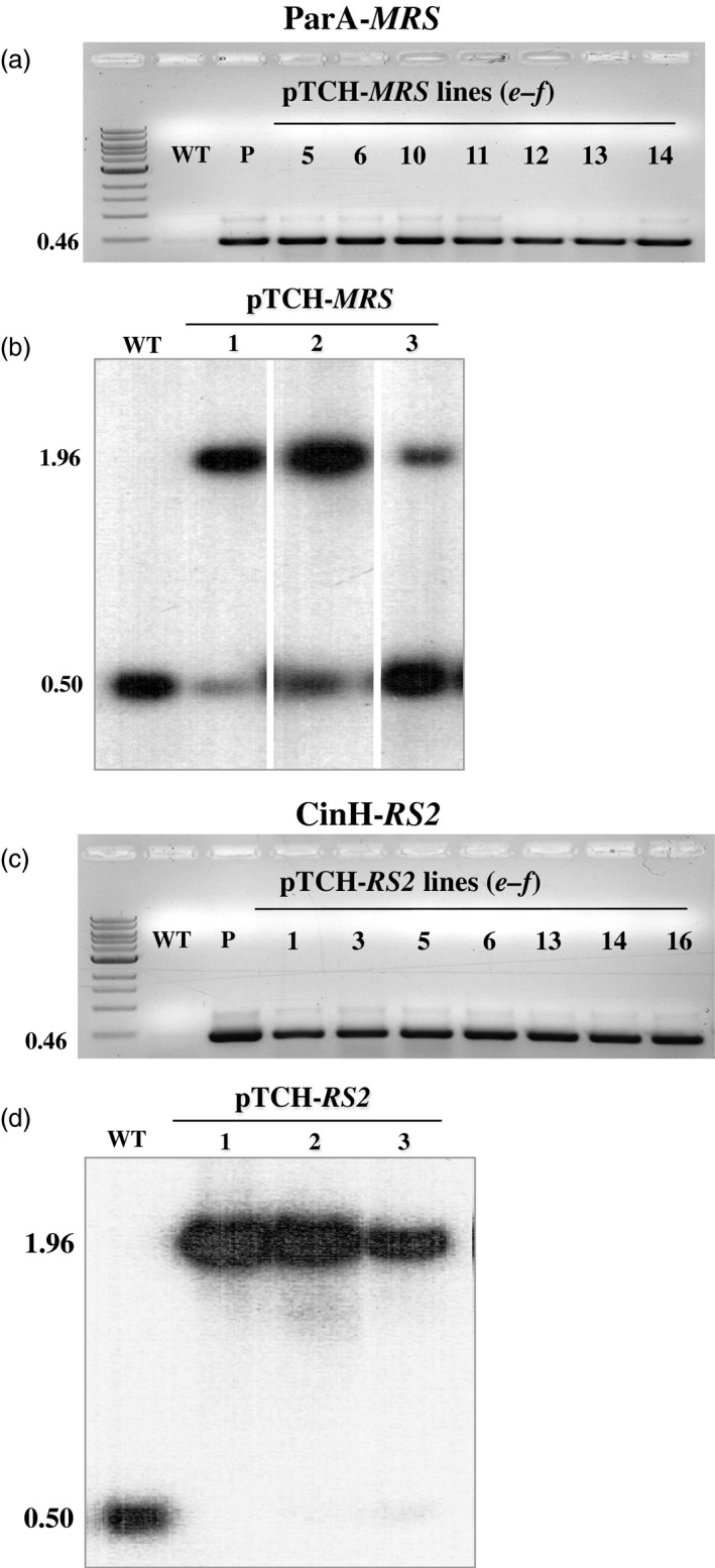
Selectable marker genes (SMGs) are necessary for selection of transgenic plants. However, once stable transformants have been identified, the marker gene is no longer needed. In this study, we demonstrate the use of the small serine recombination systems, ParA-MRS and CinH-RS2, to precisely excise a marker gene from the plastid genome of tobacco. Transplastomic plants transformed with the pTCH-MRS and pTCH-RS2 vectors, containing the visual reporter gene DsRed flanked by directly oriented MRS and RS2 recognition sites, respectively, were crossed with nuclear-genome transformed tobacco plants expressing plastid-targeted ParA and CinH recombinases, respectively. One hundred per cent of both types of F1 hybrids exhibited excision of the DsRed marker gene. PCR and Southern blot analyses of DNA from F2 plants showed that approximately 30% (CinH-RS2) or 40% (ParA-MRS) had lost the recombinase genes by segregation. The postexcision transformed plastid genomes were stable and the excision events heritable. The ParA-MRS and CinH-RS2 recombination systems will be useful tools for site-specific manipulation of the plastid genome and for generating marker-free plants, an essential step for reuse of SMG and for addressing concerns about the presence of antibiotic resistance genes in transgenic plants.

Keywords: CinH-RS2; ParA-MRS; marker excision; site-specific recombination.


Conflict of interest statement

The authors declare that they have no competing interests.

Figures

Figure 1
Figure 1
Vectors and sequences used to demonstrate precise marker gene excision from plastid DNA by ParA‐ MRS and CinH‐ RS2 systems. (a). The pTCH MRS or pTCH RS2 constructs contain either two MRS (for ParA excision) or RS2 (for CinH excision) sites (grey arrows), respectively, flanking the DsRed gene driven by the psbA promoter. The locations of Bgl II (Bg) and BamH1 (B) sites used in genomic Southern analyses are shown along with the predicted site of the fragment that hybridizes to the TRN probe (grey bar below trnI region). The sizes of the amplicons generated for each construct by primer pairs a–b and e–f (Table 1) are shown below the diagram. TrnI—chloroplast gene encoding isoleucine tRNA; trnA—chloroplast gene encoding alanine tRNA; aadA—spectinomycin 3”‐adenyltransferase gene; DsRed—red fluorescent protein gene; MRS —ParA recombinase recognition sequence; RS2—CinH recombinase recognition sequence; P 35S —Cauliflower Mosaic Virus 35S promoter. (b). p35SSTDParAo or p35SSTDCinHwt contain the recombinase expression cassettes. The size of the amplicons generated for each construct by primer pair c–d (Table 1) is shown below the diagram. ParAo—plant codon optimized ParA coding sequence; CinHwt—CinH wild‐type coding sequence; npt II—neomycin phosphotransferase II gene; STD —tobacco Rubisco stroma‐targeting domain; LB and RB—left and right, respectively, borders of T‐DNA; T–Nos 3’ terminator. (c). Excision products of site‐specific recombinase removal the DsRed gene from the plastid genome. Panels (d) and (e); Sequence of the PCR products containing ParA ( MRS ) and CinH ( RS2) recognition sites after excision—arrow between aadA and trnA genes in panel c.
Figure 2
Figure 2
Molecular analysis transplastomic plant DNA. (a) & (c). PCR analysis of spectinomycin‐resistant regenerants. DNAs were amplified using the e‐f primer pair (Table 1, Figure 1a) with the expected size of 0.46 kb shown to the right. Panel a. DNAs from WT: untransformed negative control P: positive control (pTCH MRS vector); Lanes 5, 6, 10,11, 12, 13, 14, pTCH MRS transformants; Panel c. DNAs from WT; P: positive control (pTCH RS2 vector); Lanes 1, 3, 5, 6, 13, 14 and 16 from, pTCH RS2 transformants. M, DNA ladder. (b) & (d). Southern blot analysis of PCR‐positive transplastomic plants with probe TRN (grey bar below diagram in Figure 1a). DNAs from untransformed (WT) and transplastomic plants (Lanes 1‐3) were digested with Bam HI and Bgl II. The sizes of the hybridizing fragments are shown to the left.
Figure 3
Figure 3
ParA transgenic plants and expression activity detection. (a). PCR analysis of kanamycin‐resistant regenerants. DNA from kanamycin‐resistant shoots was amplified by primer pair c–d (Figure 1b, Table 1) to give a 0.68 fragment that includes part of the 35S promoter, stroma targeting domain and part of the ParAo coding region. Amplified DNA products are from WT—untransformed tobacco negative control, P—C35SSTDParAo plasmid DNA as positive controls, and kanamycin resistant shoots (numbered lanes). (b). Schematic representative of constructions and strategy to detect recombinase activity in a transient assay. In vector pG4NG MRS GUSPlus reporter gene expression is inhibited due to the presence of a terminator‐rich ‘stuffer’ sequence in the intron (In5'tron3’). This stuffer will be excised by site‐specific recombination in cells containing ParA. The resultant pG4NG MRS exc plasmid express the GUSPlus reporter. p409sUbi—potato ubiquitin promoter; T—3'Nos terminator; In5’—maize ubiquitin first intron 5’ fragment; tron3’—maize ubiquitin first intron 3’ fragment; GUSPlus—β‐glucuronidase gene sequence; db35S‐double enhanced CaMV 35S promoter; eGFP ‐enhanced Green Fluorescent Protein gene; MRS ‐ParA recombinase recognition sequences; Amp—Ampicillin resistance gene. (c). Detection of site‐specific recombinase activity with the pG4NG MRS vector. Leaves from C35SSTDParAo transgenic plants after bombardment with the pG4NG MRS plasmid, histochemical staining to detect β‐glucuronidase activity, (blue spots) indicating recombinase‐mediated excision and gene activation. Leaves were from WT, nontransformed SR1 tobacco, the negative control, three ParAo expression transgenic plants (PAo4, PAo6 and PAo18).
Figure 4
Figure 4
CinH transgenic plants and expression activity detection. (a). PCR analysis of kanamycin‐resistant regenerants. DNA from kanamycin‐resistant shoots was amplified by primer pair c–d (Figure 1b, Table 1) to give a 0.68 or 0.52 fragment that includes part of the 35S promoter, stroma targeting domain and part of the CinH coding region. Amplified DNA products are from WT—untransformed tobacco negative control, P—C35SSTDCinH as positive controls and kanamycin resistant shoots (numbered lanes). (b). Schematic representative of constructions and strategy to detect recombinase activity in a transient assay. In vector pG4NG RS2, GUSPlus reporter gene expression is inhibited due to the presence of a terminator‐rich ‘stuffer’ sequence in the intron (In5'tron3’). This stuffer will be excised by site‐specific recombination in cells containing CinH. The resultant pG4NG RS2exc plasmid expresses the GUSPlus reporter. p409sUbi—potato ubiquitin promoter; T—3'Nos terminator; In5’—maize ubiquitin first intron 5’ fragment; tron3’—maize ubiquitin first intron 3’ fragment; GUSPlus—β‐glucuronidase gene sequence; db35S—double enhanced CaMV 35S promoter; eGFP —enhanced Green Fluorescent Protein gene; RS2—CinH recombinase recognition sequences; Amp—Ampicillin resistance gene. (c). Detection of site‐specific recombinase activity with the pG4NG RS2 vector. Leaves from C35SSTDCinH transgenic plants after bombardment with the pG4NG RS2 plasmid, histochemical staining to detect β‐glucuronidase activity, (blue spots) indicating recombinase‐mediated excision and gene activation. Leaves were from WT, nontransformed SR1 tobacco, the negative control, three CinH expression transgenic plants (CH56, CH57 and CH62).
Figure 5
Figure 5
PCR analysis to detect site‐specific excision and recombinase encoding genes in F1 progeny. PCR analysis with primer pair a–b (upper panels a, c) and c‐d (lower panels b, d) on 16 randomly kanamycin and spectinomycin selected F1 hybrid plants from crosses of THCMRS1.1 by PAo4 (panels a, b) and TCHRS1.2 by CH57 (c, d). Amplified products are from DNA from WT—nontransformed tobacco SR1, negative control; N—pTCH MRS or pTCH RS2 plasmids; E—pTCH MRS exc or pTCH RS2exc, plasmids after excision and F1 progeny of the crosses (numbered lanes). The sizes of the amplicons are indicated to the left. M: DNA size markers.
Figure 6
Figure 6
DsRed expression in leaves of pTCH MRS and pTCH RS2 transformed plants and their F1 progeny under green light excitation. (a). Fluorescence microscopic images of leaves from a TCH MRS transformed plant (nonexcised) and its F1 progeny from a cross with pollen from a ParAo recombinase expression plant (excised). (b). Bright‐field (lower) microscopic images of leaves from a TCH MRS transformed plant (nonexcised) and its F1 progeny from a cross with pollen from a ParAo recombinase expression plant (excised). (c). Fluorescence microscopic images of leaves from a TCH RS2 transformed plant (nonexcised) and its F1 progeny from a cross with pollen from a CinHwt recombinase expression plant (excised). (d). Bright‐field (lower) microscopic images of leaves from a TCH RS2 transformed plant (nonexcised) and its F1 progeny from a cross with pollen from a CinHwt recombinase expression plant (excised).
Figure 7
Figure 7
Molecular analysis for segregation of ParA site‐specific excised transplastomic DNA from the presence of the recombinase expression gene in F2 tobacco plants. (a). PCR products of DNAs from F2 progeny (numbered lanes) of the TCHMRS1.1‐PAo4 cross amplified with primers a–b . Determines the presence of excised (0.76 kb) or unexcised (2.31 kb) target. (b). PCR products of DNAs from F2 progeny (numbered lanes) of the TCHMRS1.1‐PAo4 cross amplified with primers c–d . Amplicon (0.68 kb) determines presence of parAo gene. The underlined numbers in panel b are F2 plants that contain the site‐specific excision product (panal a), but not the recombinase gene. Amplified products are from DNA from WT—nontransformed tobacco SR1, negative control; N—pTCH MRS plasmid; E—pTCH MRS exc, plasmid after excision. The sizes of the amplicons are shown to the left. M: DNA size markers. (c). Southern blot analysis of transplastomic plants with probe TRN (grey bar below diagram in Figure 1a). DNAs from untransformed (WT; 0.50 bp) and transplastomic TCHMRS1.1 plants before (N lanes; 1.96 kb) and after crossing to PAo4 (F1 lane; 2.43 kb) and the F2 progeny of the crosses (numbered lanes) were digested with Bam HI and Bgl II. The sizes in kb of the hybridizing fragments are shown to the left.
Figure 8
Figure 8
Molecular analysis for segregation of CinH site‐specific excised transplastomic DNA from the presence of the recombinase expression gene in F2 tobacco plants. (a). PCR products of DNAs from F2 progeny (numbered lanes) of the TCHRS1.2‐CH57 cross amplified with primers a–b (panel a). Determines the presence of excised (0.89 kb) or unexcised (2.32 kb) target. (b). PCR products of DNAs from F2 progeny (numbered lanes) of the TCHRS1.2‐CH57 cross amplified with primers c–d . Amplicon (0.52 kb) determine the presence of cinH gene. The underlined numbers in panel b are F2 plants that contain the site‐specific excision product (panal a), but not the recombinase gene. Amplified products from panel a are from DNA from WT—nontransformed tobacco SR1, negative control; N—pTCH RS2 plasmid; E—pTCH RS2exc, plasmid after excision. The sizes of the amplicons are shown to the left. M: DNA size markers. (c). Southern blot analysis of transplastomic plants with probe TRN (grey bar below diagram in Fig. 1a). DNAs from untransformed (WT; 0.5 kb) and transplastomic TCHRS1.2 plants before (N lanes; 1.96 kb) and after crossing to CH57 (F1 lane; 2.57 kb) and the F2 progeny of the cross (numbered lanes) were digested with Bam HI and Bgl II. The sizes in kb of the hybridizing fragments are shown to the left.

Similar articles

Cited by

References

    1. Ahmad, A. , Pereira, E.O. , Conley, A.J. , Richman, A.S. and Menassa, R. (2010) Green biofactories: recombinant protein production in plants. Recent Patents Biotechnol. 4, 242–259. - PubMed
    1. Ballester, A. , Cervera, M. and Pena, L. (2006) Efficient production of transgenic citrus plants using isopentenyl transferase positive selection and removal of the marker gene by site‐specific recombination. Plant Cell Rep. 26, 39–44. - PubMed
    1. Bevis, B.J. and Glick, B.S. (2002) Rapidly maturing variants of the Discosoma red fluorescent protein (DsRed). Nat. Biotech. 20, 83–87. - PubMed
    1. Blechl, A.E. , Lin, J.W. , Shao, M. , Thilmony, R.L. and Thomson, J.G. (2012) Bxb1 recombinase mediates site‐specific deletions in transgenic wheat. Plant Mol. Biol. Rep. 30, 1357–1366.
    1. Bock, R. (2015) Engineering plastid genomes: methods, tools, and applications in basic research and biotechnology. Annu. Rev. Plant Biol. 66, 211–241. - PubMed