Review
. 2017 Apr 19;18(4):859.
doi: 10.3390/ijms18040859.

Clarifying the Ghrelin System's Ability to Regulate Feeding Behaviours Despite Enigmatic Spatial Separation of the GHSR and Its Endogenous Ligand

Affiliations
Review

Clarifying the Ghrelin System's Ability to Regulate Feeding Behaviours Despite Enigmatic Spatial Separation of the GHSR and Its Endogenous Ligand

Alexander Edwards et al. Int J Mol Sci. .

Abstract

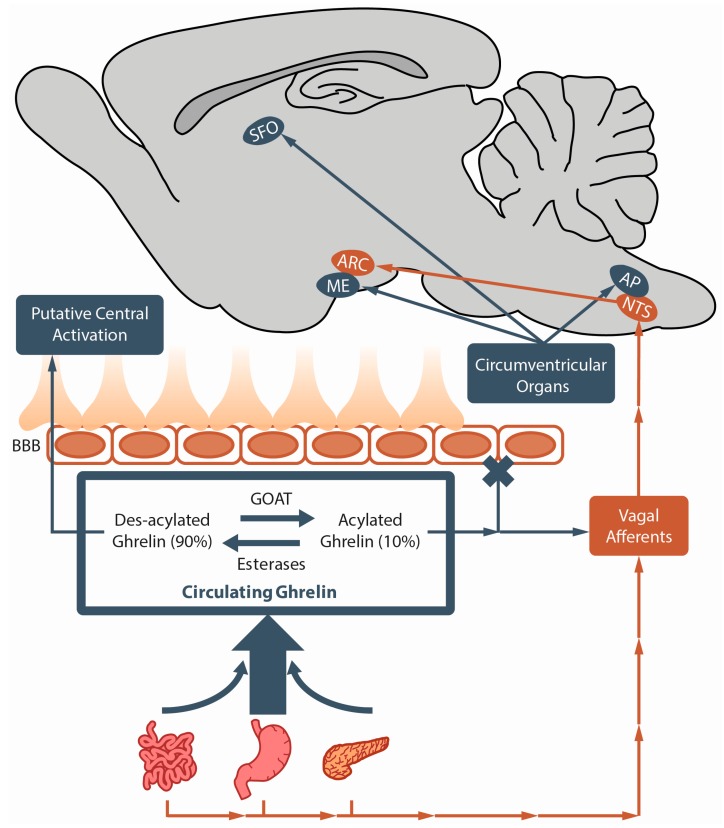
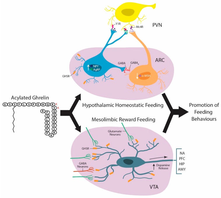
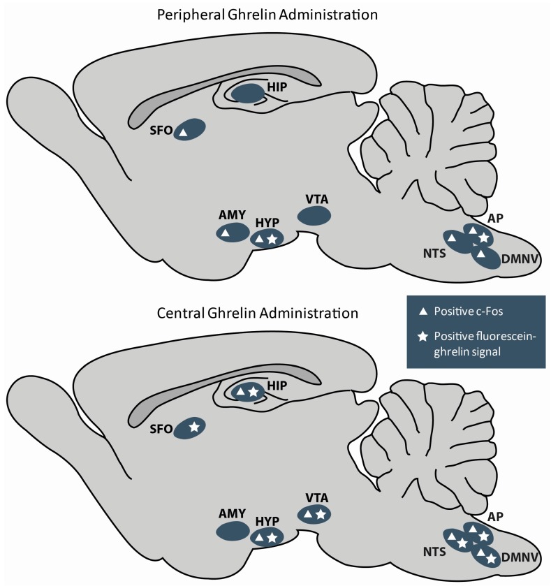
Ghrelin is a hormone predominantly produced in and secreted from the stomach. Ghrelin is involved in many physiological processes including feeding, the stress response, and in modulating learning, memory and motivational processes. Ghrelin does this by binding to its receptor, the growth hormone secretagogue receptor (GHSR), a receptor found in relatively high concentrations in hypothalamic and mesolimbic brain regions. While the feeding and metabolic effects of ghrelin can be explained by the effects of this hormone on regions of the brain that have a more permeable blood brain barrier (BBB), ghrelin produced within the periphery demonstrates a limited ability to reach extrahypothalamic regions where GHSRs are expressed. Therefore, one of the most pressing unanswered questions plaguing ghrelin research is how GHSRs, distributed in brain regions protected by the BBB, are activated despite ghrelin's predominant peripheral production and poor ability to transverse the BBB. This manuscript will describe how peripheral ghrelin activates central GHSRs to encourage feeding, and how central ghrelin synthesis and ghrelin independent activation of GHSRs may also contribute to the modulation of feeding behaviours.

Keywords: GHSR; GHSR constitutive activity; GHSR heterodimerization; blood brain barrier; central ghrelin synthesis; circumventricular organs; feeding; ghrelin; vagal afferents.


Conflict of interest statement

The authors declare no conflict of interest.

Figures

Figure 1
Figure 1
Illustration describing how peripherally produced ghrelin likely gains access to the brain. Ghrelin is primarily produced and secreted by the stomach although the small intestine and pancreas likewise produce a small quantity. Ghrelin O-acyltransferase (GOAT) converts des-acylated into its active acylated form capable of activating growth hormone secretagogue receptor (GHSRs) while esterases cleave acylated ghrelin’s O-octanoyl moiety returning it to its predominant inactive des-acylated ghrelin form (90% of total circulating ghrelin). Although des-acylated ghrelin is capable of crossing the blood brain barrier from the blood into the brain, acylated ghrelin demonstrates a very limited ability to do so (depicted by blue X). Accordingly, acylated ghrelin either stimulates GHSR on vagal afferents or bypasses the blood brain barrier (BBB) and activates GHSRs in or around circumventricular organs to convey its orexigenic effects. AP, area postrema; ARC, arcuate nucleus; ME, median eminence; NTS, nucleus tractus solitaries; SFO, subfornical organ.
Figure 2
Figure 2
Over simplified illustration depicting the two main brain regions where acylated ghrelin (human ghrelin is black amino acid sequence and red letter substitution is rat) is proposed to target to initate neurocircuits that promote feeding behaviours: the arcuate nucleus (ARC) of the hypothalamus (HYP) and the ventral tegmental area (VTA). Within the ARC, ghrelin stimulates neuropeptide Y/agouti-related peptide (NPY/AGRP) neurons by binding growth hormone secretagogue receptors (GHSRs) on their surface. Once activated theses neurons produce and release γ-aminobutyric acid (GABA) which inhibits anorectic proopiomelanocortin (POMC) neurons,decreasing the release of the anorectic peptide α-melanocyte-stimulating hormone (α-MSH). This effectively reduces the quantity of α-MSH capable of binding to satiety promoting melanocortin 4 receptors (Mc4Rs). Concurrently, activated NPY/AGRP neurons increase their production and secretion of orexigenic peptides NPY and AGRP. NPY binds to neuropeptide Y receptor type 1 (Y1R) and AGRP antagonizes the binding of α-MSH at Mc4Rs. Together the reduction in anorectic peptide and enhancement of orexigenic ones work to reduce the activity of second order anorexigenic neurons in the paraventricular nucleus (PVN) to promote homeostatic feeding behaviours. Similarly, ghrelin also stimulates VTA dopamine (DA) neurons increasing the frequency and probability of DA release from their projections in the nucleus accumbens (NA), prefrontal cortex (PFC), hippocampus (HIP), and amygdala (AMY) to encourage mesolimbic reward feeding. Ghrelin activates these VTA dopamine neurons both directly by binding to GHSR receptors located on their surface and indirectly by increasing the ratio of excitatory to inhibitory synapses contacting them.
Figure 3
Figure 3
Illustration of the feeding related brain regions that demonstrate positive c-Fos and/or fluorescein-ghrelin signals following peripheral and central ghrelin administration. Fluorescein ghrelin is a probe that has been used to determine what brain regions normal acylated ghrelin accesses. It is a fluorescently tagged (fluorescein isothiocyanate) truncated analog of the ghrelin peptide (i.e., 18 amino acids) with equivalent receptor stability, agonist activity, and binding affinity as acylated ghrelin [133]. AMY, amygdala; AP, area postrema; DMNV, dorsal motor nucleus of the vagus nerve; HIP, hippocampus; HYP, hypothalamus; NTS, nucleus tractus solitaries; SFO, subfornical organ; VTA, ventral tegmental area.

References

    1. Abizaid A., Liu Z.W., Andrews Z.B., Shanabrough M., Borok E., Elsworth J.D., Roth R.H., Sleeman M.W., Picciotto M.R., Tschöp M.H., et al. Ghrelin modulates the activity and synaptic input organization of midbrain dopamine neurons while promoting appetite. J. Clin. Investig. 2006;116:3229–3239. doi: 10.1172/JCI29867. - DOI - PMC - PubMed
    1. Asakawa A., Inui A., Kaga T., Yuzuriha H., Nagata T., Fujimiya M., Katsuura G., Makino S., Fujino M.A., Kasuga M. A role of ghrelin in neuroendocrine and behavioral responses to stress in mice. Neuroendocrinology. 2001;74:143–147. doi: 10.1159/000054680. - DOI - PubMed
    1. Diano S., Farr S.A., Benoit S.C., McNay E.C., da Silva I., Horvath B., Gaskin F.S., Nonaka N., Jaeger L.B., Banks W.A., et al. Ghrelin controls hippocampal spine synapse density and memory performance. Nat. Neurosci. 2006;9:381–388. doi: 10.1038/nn1656. - DOI - PubMed
    1. Howard A.D., Feighner S.D., Cully D.F., Arena J.P., Paul A., Rosenblum C.I., Hamelin M., Hreniuk D.L., Palyha O.C., Hamelin M., et al. A receptor in pituitary and hypothalamus that functions in growth hormone release. Science. 1996;273:974–977. doi: 10.1126/science.273.5277.974. - DOI - PubMed
    1. Lutter M., Sakata I., Osborne-Lawrence S., Rovinsky S.A., Anderson J.G., Jung S., Birnbaum S., Yanagisawa M., Elmquist J.K., Nestler E.J., et al. The orexigenic hormone ghrelin defends against depressive symptoms of chronic stress. Nat. Neurosci. 2008;11:752–753. doi: 10.1038/nn.2139. - DOI - PMC - PubMed

LinkOut - more resources